. 2013 Aug 9;2(10):990-7.
doi: 10.1242/bio.20135041. eCollection 2013.

The role of Tal2 and Tal1 in the differentiation of midbrain GABAergic neuron precursors

Affiliations

The role of Tal2 and Tal1 in the differentiation of midbrain GABAergic neuron precursors

Kaia Achim et al. Biol Open. .

Abstract

Midbrain- and hindbrain-derived GABAergic interneurons are critical for regulation of sleep, respiratory, sensory-motor and motivational processes, and they are implicated in human neurological disorders. However, the precise mechanisms that underlie generation of GABAergic neuron diversity in the midbrain-hindbrain region are poorly understood. Here, we show unique and overlapping requirements for the related bHLH proteins Tal1 and Tal2 in GABAergic neurogenesis in the midbrain. We show that Tal2 and Tal1 are specifically and sequentially activated during midbrain GABAergic neurogenesis. Similar to Gata2, a post-mitotic selector of the midbrain GABAergic neuron identity, Tal2 expression is activated very early during GABAergic neuron differentiation. Although the expression of Tal2 and Gata2 genes are independent of each other, Tal2 is important for normal midbrain GABAergic neurogenesis, possibly as a partner of Gata2. In the absence of Tal2, the majority of midbrain GABAergic neurons switch to a glutamatergic-like phenotype. In contrast, Tal1 expression is activated in a Gata2 and Tal2 dependent fashion in the more mature midbrain GABAergic neuron precursors, but Tal1 alone is not required for GABAergic neuron differentiation from the midbrain neuroepithelium. However, inactivation of both Tal2 and Tal1 in the developing midbrain suggests that the two factors co-operate to guide GABAergic neuron differentiation in a specific ventro-lateral midbrain domain. The observed similarities and differences between Tal1/Tal2 and Gata2 mutants suggest both co-operative and unique roles for these factors in determination of midbrain GABAergic neuron identities.

Keywords: Brain development; Dopaminergic neuron; GABAergic neuron; Gata; Hindbrain; Midbrain; Mouse; Neurogenesis; Rhombomere 1; Scl; Substantia nigra pars reticulata (SNpr); Tal; Transcription factor; Ventral tegmental area (VTA).


Conflict of interest statement

Competing interests: The authors have no competing interests to declare.

Figures

Fig. 1.
Fig. 1.. Expression of Tal and Gata factors in the wild-type and Tal1cko midbrain.
ISH analysis of Gad1, Tal1, Tal2, Gata2, and Gata3 expression on adjacent coronal sections of WT (A–J) and Tal1cko (K–O) midbrains at E10.5–E11.5. Radioactive ISH in panels A–E, non-radioactive in the others. The development of GABAergic neurons remains unaffected in the Tal1cko midbrain. Tal1flox probe (G,L) was designed to recognize the region between LoxP sites in the conditional allele and therefore the loss of signal in the Tal1cko midbrain demonstrates efficient inactivation of the Tal1 gene.
Fig. 2.
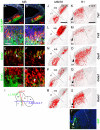
Fig. 2.. Expression of Tal and Gata factors in the developing midbrain and r1.
(A–H) Co-expression of Tal2 and Tal1 (ISH) with Gad67-driven GFP, Gata2, and Gata3 in coronal sections of E11.5 midbrains. Epifluorescence images in (A,B,E–H), confocal images in (C,D). (I) The planes of sectioning. (J–S) Radioactive ISH with the probes indicated adjacent coronal sections from E11.5 midbrain–r1 (J,L,N,P,R). The dashed line indicates the midbrain–r1 border, defined by the anti-Otx2 staining on an adjacent section (not shown). (K,M,O,Q,S) Radioactive ISH with the probes indicated on parallel transverse sections from E12.5 rhombomere 1 area. The dashed lines mark the ventral and dorsal borders of Nkx6.1 expression domain in r1 (not shown). The serotonergic domain (SA) was detected using with an anti-5HT antibody on a parallel section (T). r1, rhombomere 1, mb, midbrain. White arrowheads in C–H indicate co-expression, arrowheads in K–T point to serotonergic neurons. Scale bars: 100 µm (A,J), 25 µm (C), 200 µm (K).
Fig. 3.
Fig. 3.. Expression of Tal factors in the intermediate and mantle zones of the midbrain.
(A,B) Fluorescent ISH with Tal1 and Tal2 probes combined with p57 IHC on E11.5 ventral midbrain. The positions of three zones were deduced from the p57+ cells, which mark the intermediate zone. (C–E) Fluorescent ISH with Tal1 and Tal2, and IHC with a pan-neuronal marker HuC/D on E11.5 ventral midbrain. (F,G) Fluorescent ISH with Tal2 and IHC with ventricular zone marker Sox2 on E11.5 wild-type and Gata2cko ventral midbrain, with domain borders indicated. The dashed line visualizes the border between ventricular zone and mantle zone, based on Sox2 staining. VZ, ventricular zone; IZ, intermediate zone; MZ, mantle zone; mb, midbrain; r1, rhombomere 1. Scale bars: 25 µm (A–E), 100 µm (F,G).
Fig. 4.
Fig. 4.. Tal1 and Tal2 expression in the Gata2 mutants.
ISH analysis of Gad1 (A,B,G,H), Tal1 (C,D,I,J) and Tal2 (E,F,K,L) expression in wild-type (WT) and Gata2cko embryos at E12.5 and at E15.5. Note that the expression of Tal2 in the r1 is below the detection level by these non-radioactive ISH experiments. The dashed line indicates the midbrain/r1 border, detected by Otx2 staining on a parallel section (data not shown). Arrowheads point to the SNpr, defined by TH immunostaining on adjacent sections (not shown). Arrows point to Tal2 expression domain in the dorsal midbrain (close up images in insets, K′ and L′). mb, midbrain; r1, rhombomere 1; PAG, periaqueductal grey; mRF, midbrain reticular formation; SNpr, substantia nigra pars reticulata, SC, superior colliculi. Scale bars: 200 µm.
Fig. 5.
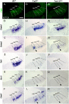
Fig. 5.. GABAergic gene expression in Tal2 and Tal1/2dko mutant midbrains.
Expression of Helt (IHC), Gata2, Gata3, Tal1, Six3 and Gad1 (ISH) on coronal sections of E11.5 wild-type (A–F), Tal2ko (G–L) and Tal1/2dko (M–R) midbrains. The dashed lines indicate the borders of midbrain m3–m5 domains, defined by Helt and Nkx2.2 stainings on parallel sections (not shown). The arrowheads point to the m5 domain, where fate transformation appears to be incomplete. Scale bar: 100 µm.
Fig. 6.
Fig. 6.. Glutamatergic gene expression in Tal2 and Tal1/2dko mutant midbrains.
Expression of Nkx6.1 (A,D,G), Pax6 (B,E,H) and Scl17a6 (C,F,I) on coronal sections of E11.5 wild-type (A–C), Tal2ko (D–F) and Tal1/2dko (G–I) midbrains. The dashed lines indicating the borders of m3–m5 domains were defined by Helt and Nkx2.2 stainings on parallel sections (not shown). Arrowheads point to ectopic expression. Arrows in panels D and G indicate areas where ectopic Pax6 expression is seen on a parallel section. Scale bars: 100 µm.
Fig. 7.
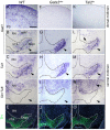
Fig. 7.. GABAergic and glutamatergic marker gene expression in the Gata2cko and Tal2ko perinatal brain.
The expression of Gad1, Tal1 and Slc17a6 (ISH) was analyzed on coronal sections through the WT (A–D), Gata2cko (F–I), and Tal2ko (K–N) midbrains at P0. TH IHC on adjacent sections (E,J,O) was used as a landmark to identify the midbrain region and define the SN/VTA nuclei. The arrowheads point to SN, the arrow to mRF. Scale bars: 200 µm.
Fig. 8.
Fig. 8.. Model of Gata/Tal terminal selector complex and its variants in the midbrain.
A schematic cross-section of E12.5 midbrain, with the domains generating GABAergic neurons indicated in green. Both Gata2 and Tal2 expression is initiated after cell cycle exit. Coincident expression of Gata2 and Tal2 leads to formation of a terminal selector complex crucial for the correct GABAergic identity development in m1–4d. In m5, Tal1 is also required. VZ, ventricular zone; IZ, intermediate zone; MZ, mantle zone; SA, serotonergic domain.

References

    1. Achim K., Peltopuro P., Lahti L., Li J., Salminen M., Partanen J. (2012). Distinct developmental origins and regulatory mechanisms for GABAergic neurons associated with dopaminergic nuclei in the ventral mesodiencephalic region. Development 139, 2360–2370 10.1242/dev.076380 - DOI - PubMed
    1. Bradley C. K., Takano E. A., Hall M. A., Göthert J. R., Harvey A. R., Begley C. G., van Eekelen J. A. (2006). The essential haematopoietic transcription factor Scl is also critical for neuronal development. Eur. J. Neurosci. 23, 1677–1689 10.1111/j.1460-9568.2006.04712.x - DOI - PubMed
    1. Bucher K., Sofroniew M. V., Pannell R., Impey H., Smith A. J., Torres E. M., Dunnett S. B., Jin Y., Baer R., Rabbitts T. H. (2000). The T cell oncogene Tal2 is necessary for normal development of the mouse brain. Dev. Biol. 227, 533–544 10.1006/dbio.2000.9920 - DOI - PubMed
    1. Cantor A. B., Orkin S. H. (2002). Transcriptional regulation of erythropoiesis: an affair involving multiple partners. Oncogene 21, 3368–3376 10.1038/sj.onc.1205326 - DOI - PubMed
    1. Cohen J. Y., Haesler S., Vong L., Lowell B. B., Uchida N. (2012). Neuron-type-specific signals for reward and punishment in the ventral tegmental area. Nature 482, 85–88 10.1038/nature10754 - DOI - PMC - PubMed