. 2014 Mar;20(5-6):1027-37.
doi: 10.1089/ten.TEA.2013.0111. Epub 2013 Dec 11.

Long-term characterization of axon regeneration and matrix changes using multiple channel bridges for spinal cord regeneration

Affiliations

Long-term characterization of axon regeneration and matrix changes using multiple channel bridges for spinal cord regeneration

Hannah M Tuinstra et al. Tissue Eng Part A. 2014 Mar.

Abstract

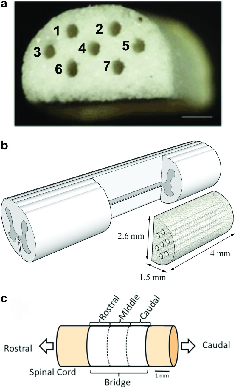
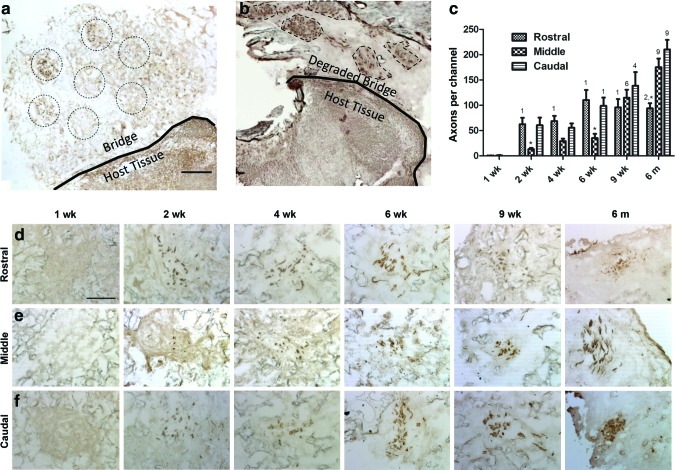
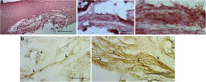
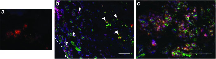
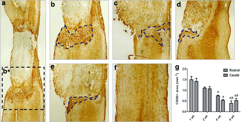
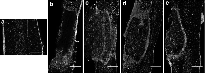
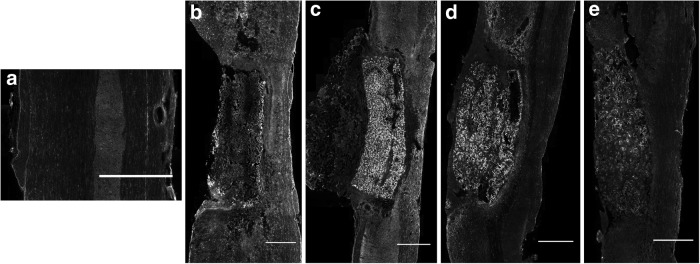
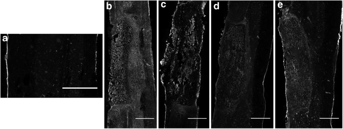
Spinal cord injury (SCI) results in loss of sensory and motor function below the level of injury and has limited available therapies. The host response to SCI is typified by limited endogenous repair, and biomaterial bridges offer the potential to alter the microenvironment to promote regeneration. Porous multiple channel bridges implanted into the injury provide stability to limit secondary damage and support cell infiltration that limits cavity formation. At the same time, the channels provide a path that physically directs axon growth across the injury. Using a rat spinal cord hemisection injury model, we investigated the dynamics of axon growth, myelination, and scar formation within and around the bridge in vivo for 6 months, at which time the bridge has fully degraded. Axons grew into and through the channels, and the density increased overtime, resulting in the greatest axon density at 6 months postimplantation, despite complete degradation of the bridge by that time point. Furthermore, the persistence of these axons contrasts with reports of axonal dieback in other models and is consistent with axon stability resulting from some degree of connectivity. Immunostaining of axons revealed both motor and sensory origins of the axons found in the channels of the bridge. Extensive myelination was observed throughout the bridge at 6 months, with centrally located and peripheral channels seemingly myelinated by oligodendrocytes and Schwann cells, respectively. Chondroitin sulfate proteoglycan deposition was restricted to the edges of the bridge, was greatest at 1 week, and significantly decreased by 6 weeks. The dynamics of collagen I and IV, laminin, and fibronectin deposition varied with time. These studies demonstrate that the bridge structure can support substantial long-term axon growth and myelination with limited scar formation.


Figures

<b>FIG. 1.</b>
FIG. 1.
Multiple channel bridges for spinal cord regeneration. (a) Photomicrograph of a multiple channel bridge showing seven channels, each 250 μm in diameter. Scale bar is 500 μm. (b) Schematic representation of PLG bridge implantation in a spinal cord hemisection. (c) Schematic representation of the regions in which the bridge was divided for analysis. Rostral analysis was performed at 300 μm, middle at 2000 μm, and caudal at 3500 μm from the rostral edge of the bridge/tissue boundary. Color images available online at www.liebertpub.com/tea
<b>FIG. 2.</b>
FIG. 2.
Time course of axon growth in channels of the bridge. (a) NF200-stained cross section of the bridge at 9 weeks with seven channels indicated by dashed circles (b) NF200-stained cross section of the injury site at >6 months with a fully degraded bridge and at least five remaining neurofilament bundles outlined with dashes. Axons were stained at 1, 2, 4, 6, 9 weeks, and 6 months at (c) rostral (300 μm), (d) middle (2000 μm), and (e) caudal (3500 μm) regions of the bridge. (f ) Quantification of axonal regeneration as a function of time and position within the bridges was performed by counting the number of NF200-positive axons inside the channels. Axon density was a function of time (ANOVA, F=56.8, p<0.0001) and location within the bridge (ANOVA, F=9.8, p<0.0001). Slides analyzed were selected from the NF200-stained cross sections as in (c–e). Statistical analysis was carried out by an ANOVA with Bonferroni post-test with a p<0.05 found to be significantly different. “*,” significant difference compared to other locations at the same time point; “1, 2, 4, 6, 9,” significant difference compared to 1, 2, 4, 6, and 9 weeks (respectively) and previous at the same location. Scale bar in (a, b) is 250 μm and in (c–e) is 50 μm. Color images available online at www.liebertpub.com/tea
<b>FIG. 3.</b>
FIG. 3.
Motor and sensory axons within channels of the bridge at 8 weeks. (a) ChAT-positive axons (red) within the channels of the bridge indicated by the arrowheads. Scale bars are 1000, 100, and 50 mm from left to right. (b) CGRP-positive axons (brown) within the channels of a bridge indicated by the arrows. Scale bars are 50 mm. Color images available online at www.liebertpub.com/tea
<b>FIG. 4.</b>
FIG. 4.
Myelination of axons within the injury site overtime. Transverse tissue sections were stained with MBP (red), P0 (blue), and NF200 (green). (a) Cross section of a channel at 6 weeks stained positive for MBP but no P0 myelin. (b, c) At 6 months, extensive myelination was observed throughout the injury site. Co-localization of P0 and MBP myelin was observed around the outer curved surface of the bridge, indicated by open arrowheads in (b) and throughout in (c). While mostly MBP, only myelin was observed toward the midline and center channels (numbered 4, 6, and 7 in Fig. 1a) indicated by the closed arrowheads in (b). Scale bar is 100 μm. Color images available online at www.liebertpub.com/tea
<b>FIG. 5.</b>
FIG. 5.
Chondroitin sulfate proteoglycans (CSPG) staining over time. (a) CS-56-stained tissue section at 1 week with location of inset b* indicated by dashed lines. (b–e) Caudal sections at 1, 2, 6, and 9 weeks postimplantation with positive staining indicated by dashed lines. (f ) Uninjured cord-negative control. (g) Quantification of the CS-56-positive area directly rostral and caudal to the bridge. “a,” statistically different from 1 week at the same location; “b,” statistically different from 2 weeks at the same location, p<0.05. Color images available online at www.liebertpub.com/tea
<b>FIG. 6.</b>
FIG. 6.
Collagen IV-stained tissue sections of uninjured cord (a) and 1, 2, 6, and 9 weeks postimplantation (b–e). Scale bar is 1 mm.
<b>FIG. 7.</b>
FIG. 7.
Laminin-stained tissue sections of uninjured cord (a) and 1, 2, 6, and 9 weeks postimplantation (b–e). Scale bar is 1 mm.
<b>FIG. 8.</b>
FIG. 8.
Collagen I-stained tissue sections of uninjured cord (a) and 1, 2, 6, and 9 weeks postimplantation (b–e). Scale bar is 1 mm.
<b>FIG. 9.</b>
FIG. 9.
Fibronectin-stained tissue sections of uninjured cord (a) and 1, 2, 6, and 9 weeks postimplantation (b–e). Scale bar is 1 mm.

Similar articles

Cited by

References

    1. Richardson P.M., McGuinness U.M., and Aguayo A.J.Axons from CNS neurons regenerate into PNS grafts. Nature 284,264, 1980 - PubMed
    1. David S., and Aguayo A.J.Axonal elongation into peripheral nervous system “bridges” after central nervous system injury in adult rats. Science 214,931, 1981 - PubMed
    1. Geller H.M., and Fawcett J.W.Building a bridge: engineering spinal cord repair. Exp Neurol 174,125, 2002 - PubMed
    1. Joosten E.A.Biodegradable biomatrices and bridging the injured spinal cord: the corticospinal tract as a proof of principle. Cell Tissue Res 349,375, 2012 - PMC - PubMed
    1. Wang M., Zhai P., Chen X., Schreyer D.J., Sun X., and Cui F.Bioengineered scaffolds for spinal cord repair. Tissue Eng Part B Rev 17,177, 2011 - PubMed

Publication types

MeSH terms