. 2013 Nov 26;52(47):8430-41.
doi: 10.1021/bi4010914. Epub 2013 Nov 14.

Pulsed EPR study of amino acid and tetrahydropterin binding in a tyrosine hydroxylase nitric oxide complex: evidence for substrate rearrangements in the formation of the oxygen-reactive complex

Affiliations

Pulsed EPR study of amino acid and tetrahydropterin binding in a tyrosine hydroxylase nitric oxide complex: evidence for substrate rearrangements in the formation of the oxygen-reactive complex

Matthew D Krzyaniak et al. Biochemistry. .

Abstract

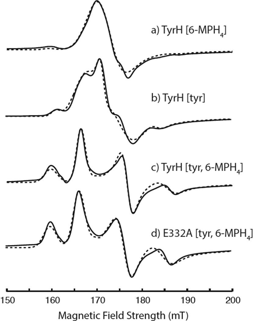
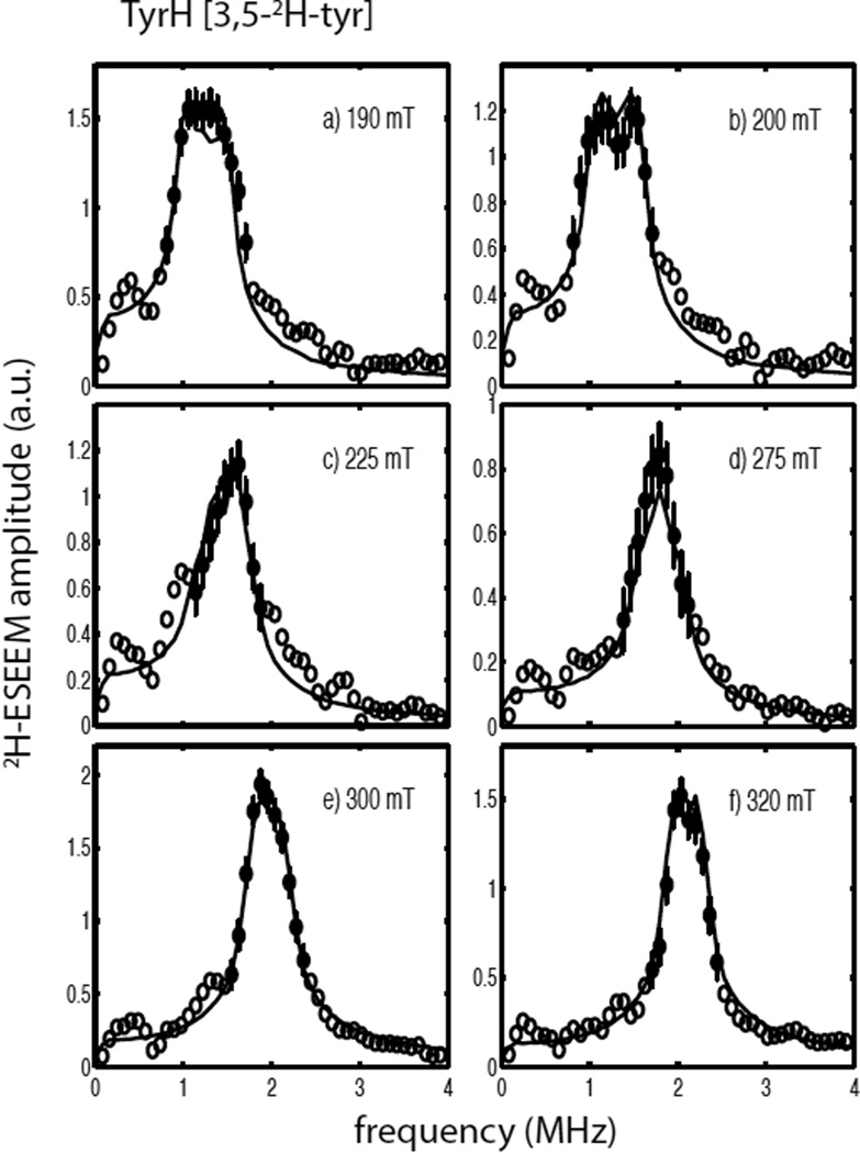
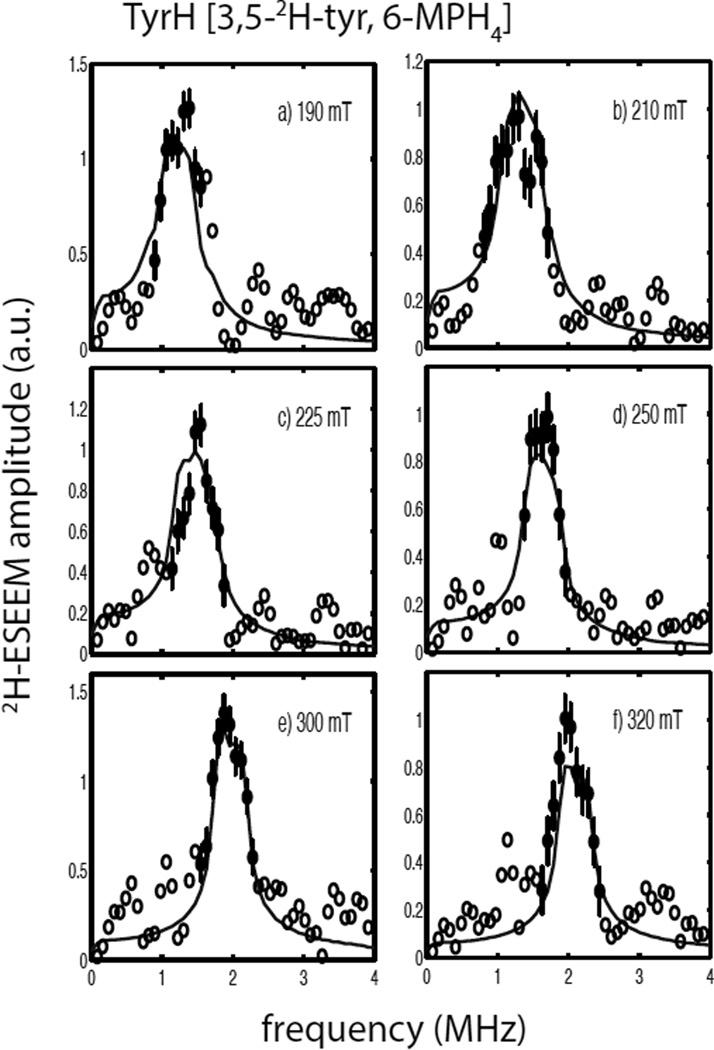
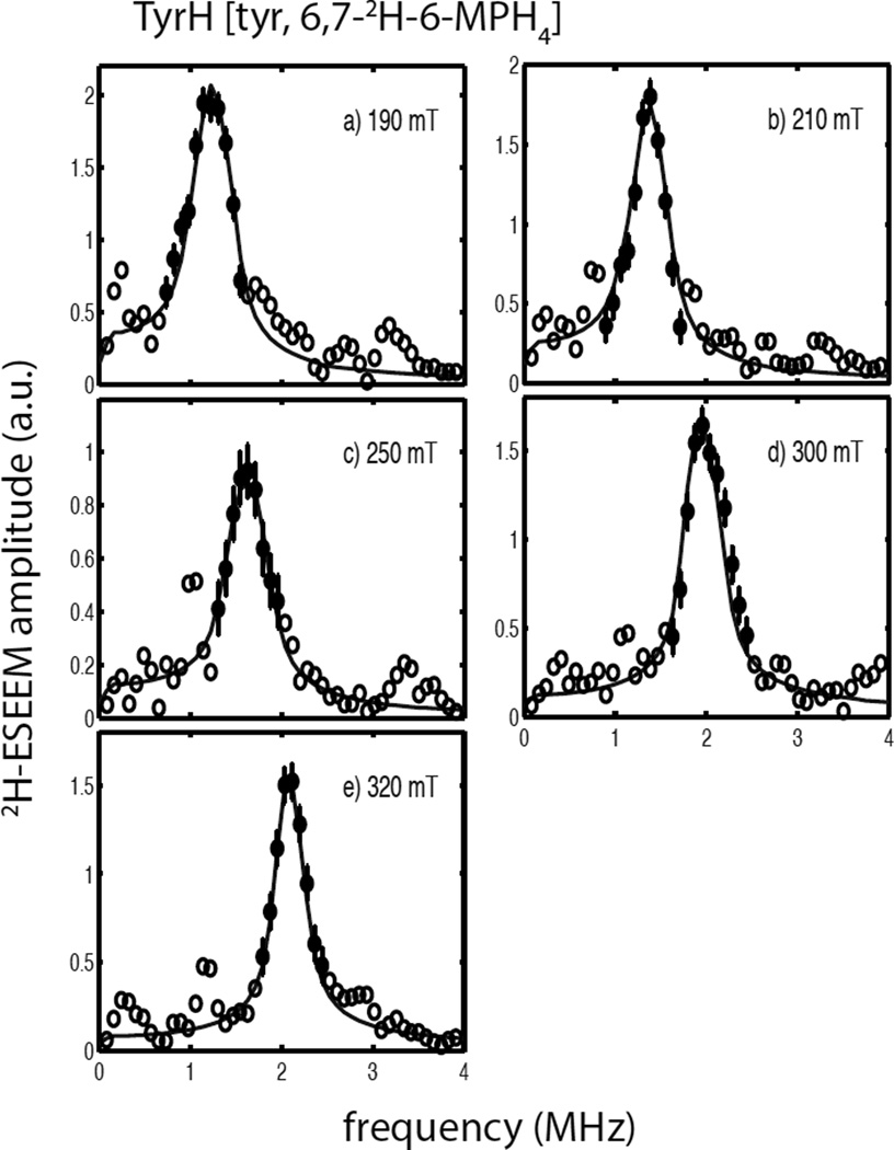
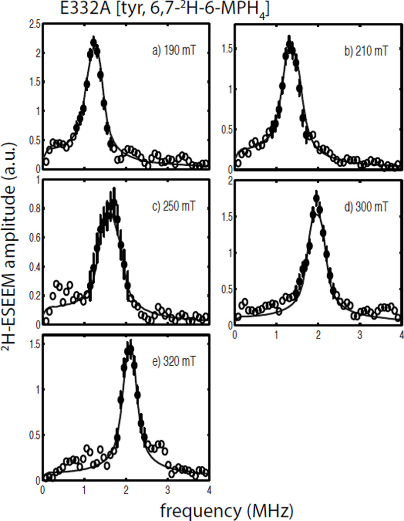
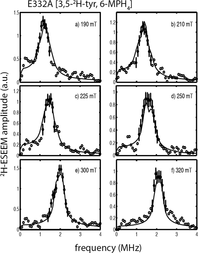
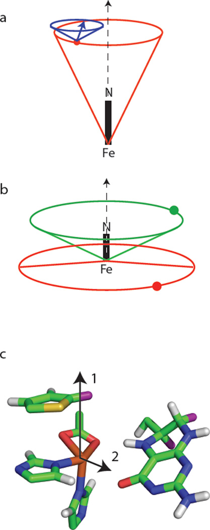
Tyrosine hydroxylase is a nonheme iron enzyme found in the nervous system that catalyzes the hydroxylation of tyrosine to form l-3,4-dihydroxyphenylalanine, the rate-limiting step in the biosynthesis of the catecholamine neurotransmitters. Catalysis requires the binding of three substrates: tyrosine, tetrahydrobiopterin, and molecular oxygen. We have used nitric oxide as an O₂ surrogate to poise Fe(II) at the catalytic site in an S = 3/2, {FeNO}⁷ form amenable to EPR spectroscopy. ²H-electron spin echo envelope modulation was then used to measure the distance and orientation of specifically deuterated substrate tyrosine and cofactor 6-methyltetrahydropterin with respect to the magnetic axes of the {FeNO}⁷ paramagnetic center. Our results show that the addition of tyrosine triggers a conformational change in the enzyme that reduces the distance from the {FeNO}⁷ center to the closest deuteron on 6,7-²H-6-methyltetrahydropterin from >5.9 Å to 4.4 ± 0.2 Å. Conversely, the addition of 6-methyltetrahydropterin to enzyme samples treated with 3,5-²H-tyrosine resulted in reorientation of the magnetic axes of the S = 3/2, {FeNO}⁷ center with respect to the deuterated substrate. Taken together, these results show that the coordination of both substrate and cofactor direct the coordination of NO to Fe(II) at the active site. Parallel studies of a quaternary complex of an uncoupled tyrosine hydroxylase variant, E332A, show no change in the hyperfine coupling to substrate tyrosine and cofactor 6-methyltetrahydropterin. Our results are discussed in the context of previous spectroscopic and X-ray crystallographic studies done on tyrosine hydroxylase and phenylalanine hydroxylase.


Figures

Figure 1
Figure 1
The g = 4 region of cw-EPR spectra collected for {FeNO}7 derivatives of TyrH treated with (a) 6-MPH4, (b) tyr, (c) 6-MPH4 plus tyr, (d) the E332A variant of TyrH treated with 6-MPH4 plus tyr. The solid lines are the experimental spectra and the simulations performed using the parameters in Table 1 are shown with dashed lines. The experimental data were collected under the following conditions: microwave frequency, 9.68 GHz; microwave power, 0.0063 mW; field modulation amplitude, 0.8 mT; 10 kHz modulation frequency; time constant, 40 msec; sample temperature, 4.0 K.
Figure 2
Figure 2
(a) 3-pulse ESEEM data collected for TyrH/NO/tyrosine (solid line) and TyrH/NO/ 3,5-2H- tyrosine (dashed line) under the following conditions: field strength, 300 mT; microwave frequency, 9.684 GHz; tau, 156 ns; sample temperature, 4.0 K. (b) The ratio of the two time domain data sets shown in (a).
Figure 3
Figure 3
2H-ESEEM spectra (open and filled circles) obtained by Fourier transformation of ESEEM data from the ternary complexes of TyrH/NO/3,5-2H- tyrosine divided by TyrH/NO/tyr. Data were collected at 9.684 GHz using the following field positions and tau values a) 190 mT, 124 ns; b) 200 mT, 116 ns; c) 225 mT, 104 ns; d) 275 mT, 84 ns; e) 300 mT, 156 ns; and f) 320 mT, 148 ns. The solid lines in each frame are best fit ESEEM simulations to the data points represented by the filled circles with error bars using the following spin Hamiltonian parameters: gn = 0.8574, T = 0.18 MHz, βhf = 26°, γhf = 0°, e2qQ/h = 0.27 MHz, βnqi = 62° and γnqi = 38°.
Figure 4
Figure 4
2H-ESEEM spectra (open and filled circles) obtained by Fourier transformation of ESEEM data from the quaternary complexes of TyrH/NO/3,5-2H- tyr/6-MPH4 divided by TyrH/ NO/tyr/6-MPH4. Data were collected at 9.68 GHz using the following field positions and tau values a) 190 mT, 124 ns; b) 210 mT, 112 ns; c) 225 mT, 104 ns; d) 250 mT, 92 ns; e) 300 mT, 156 ns; and f) 320 mT, 148 ns. The solid lines in each frame are best fit ESEEM simulations to the data points represented by the filled circles with error bars using the following spin Hamiltonian parameters: gn = 0.8574, T = 0.12 MHz, βhf = 94°, γhf = 20°, e2qQ/h = 0.34 MHz, βnqi = 71° and γnqi = 40°.
Figure 5
Figure 5
3-pulse 2H-ESEEM spectra obtained for the ternary complex TyrH/NO/6,7-2H-6-MPH4 (dashed line) and the quaternary complex TyrH/NO/tyr/6,7-2H-6-MPH4 (solid line) at (a) 178 mT, τ = 132 ns and (b) 320 mT, τ = 148 ns.
Figure 6
Figure 6
2H-ESEEM spectra (open and filled circles) obtained by Fourier transformation of ESEEM data from the ternary complexes of TyrH/NO/tyrosine/6,7-2H-6-MPH4 divided by TyrH/ NO/tyr/6-MPH4. Data were collected at 9.68 GHz using the following field positions and tau values a) 190 mT, 124 ns; b) 210 mT, 112 ns; c) 250 mT, 92 ns; d) 300 mT, 156 ns; and e) 320 mT, 148 ns. The solid lines in each frame are best fit ESEEM simulations to the data points represented by the filled circles with error bars using the following spin Hamiltonian parameters: gn = 0.8574, T = 0.14 MHz, βhf = 66°, γhf = 0°, e2qQ/h = 0.22 MHz, βnqi = 70° and γnqi = 26°.
Figure 7
Figure 7
2H-ESEEM spectra (open and filled circles) obtained by Fourier transformation of ESEEM data from the ternary complexes of E332A/NO/tyrosine/6,7-2H-6-MPH4 divided by E332A/NO/tyr/6-MPH4. Data were collected at 9.68 GHz using the following field positions and tau values a) 190 mT, 124 ns; b) 210 mT, 112 ns; c) 250 mT, 92 ns; d) 300 mT, 156 ns; and e) 320 mT, 148 ns. The solid lines in each frame are best fit ESEEM simulations to the data points represented by the filled circles with error bars using the following spin Hamiltonian parameters: gn = 0.8574, T = 0.14 MHz, βhf = 65°, γhf = 0°, e2qQ/h = 0.20 MHz, γnqi = 66° and γnqi = 18°.
Figure 8
Figure 8
2H-ESEEM spectra (open and filled circles) obtained by Fourier transformation of ESEEM data from the ternary complexes of E332A/NO/3,5-2H-tyr/6-MPH4 divided by E332A/ NO/3,5-2H-tyr/6-MPH4. Data were collected at 9.68 GHz using the following field positions and tau values a) 190 mT, 124 ns; b) 210 mT, 112 ns; c) 225 mT, 104 ns; d) 250 mT, 92 ns; e) 300 mT, 156 ns; and f) 320 mT, 148 ns. The solid lines in each frame are best fit ESEEM simulations to the data points represented by the filled circles with error bars using the following spin Hamiltonian parameters: gn = 0.8574, T = 0.12 MHz, βhf = 89°, γhf = 20°, e2qQ/h = 0.29 MHz, βnqi = 44° and γnqi = 34°.
Figure 9
Figure 9
Cone diagrams for schematic representation of the 2H spin Hamiltonian parameters given in Table 2 and obtained for (a) the ternary complex of TyrH/NO/3,5-2H-tyr; (b) the quaternary complexes of TyrH/NO/3,5-2H-tyr/6-MPH4 (red) and TyrH/NO/tyr/6,7-2H-6-MPH4 (green). Figure (c) shows a stick diagram of the catalytic site of PheH crystalized with thienylalanine and BH4 (taken from PDB file: 1MMK). The vectors show possible orientations for the Fe-NO bond axis.
Scheme 1
Scheme 1
Hydroxylation reactions catalyzed by Tyrosine Hydroxylase

References

    1. Fitzpatrick PF. Tyrosine Hydroxylase. Annu. Rev. Biochem. 1999;68:355–381. - PubMed
    1. Ludecke B, Knappskog PM, Clayton PT, Surtees RAH, Clelland JD, Heales SJR, Brand MP, Batholome K, Flatmark T. Recessively inherited L-DOPA responsive Parkinsonism in infancy caused by point mutation of the tyrosine hydroxylase gene. Hum. Mol. Genet. 1996;5:1023–1028. - PubMed
    1. Ludecke B, Dworniczak B, Bartholome K. A point mutation in the tyrosine hydroxylase gene associated with Segawa's Syndrome. Hum. Genet. 1995;95:123–125. - PubMed
    1. Smyth C, Kalsi G, Brynjolfsson J, O'Neill J, Curtis D, Rifkin L, Maloney E, Murphy P, Sherrington R, Petursson H, Gurling H. Further tests for linkage of bipolar affective disorder to the tyrosine hydroxylase gene locus. Am. J. Psychiatry. 1996;153:271–274. - PubMed
    1. Fitzpatrick PF. Mechanism of Aromatic Amino Acid Hydroxylation. Biochemistry. 2003;42:14083–14091. - PMC - PubMed

Publication types

MeSH terms