. 2013 Nov 27;32(23):3055-65.
doi: 10.1038/emboj.2013.232. Epub 2013 Oct 29.

Interferon lambda 4 signals via the IFNλ receptor to regulate antiviral activity against HCV and coronaviruses

Affiliations

Interferon lambda 4 signals via the IFNλ receptor to regulate antiviral activity against HCV and coronaviruses

Ole J Hamming et al. EMBO J. .

Abstract

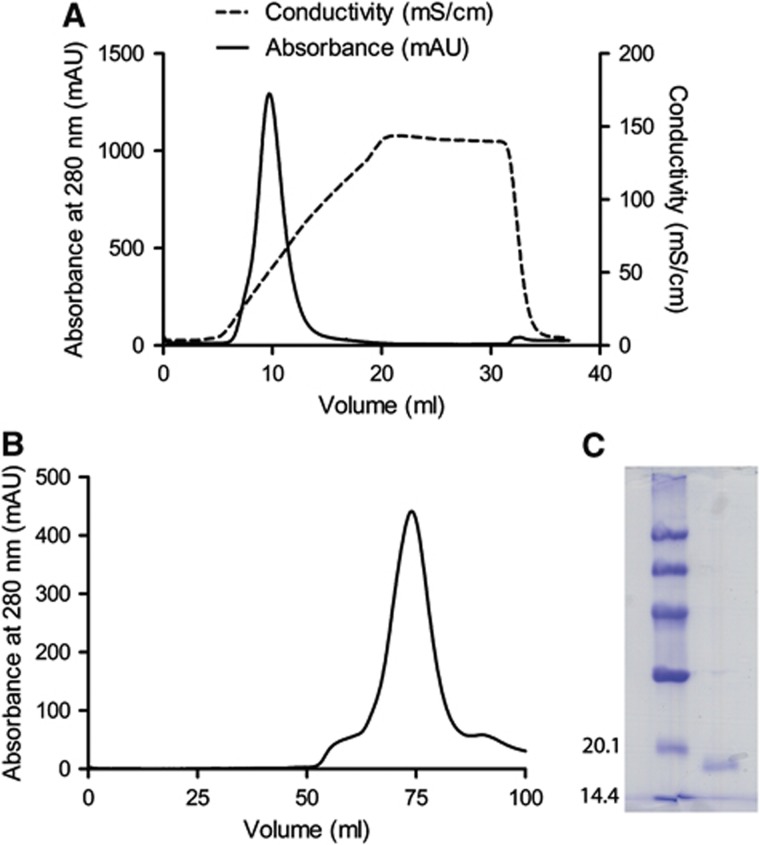
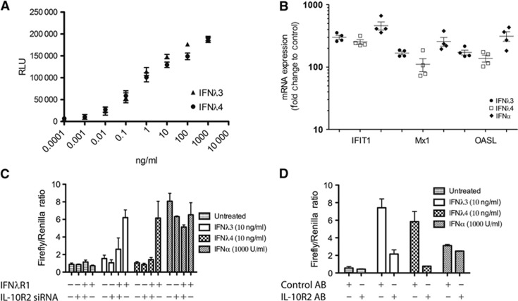
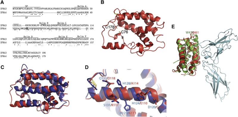
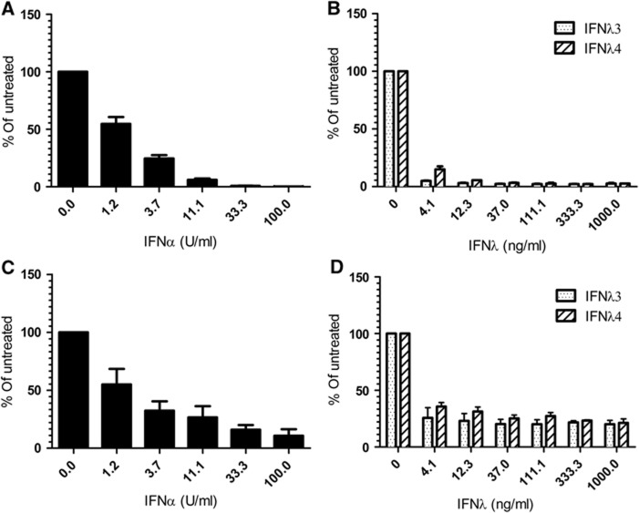
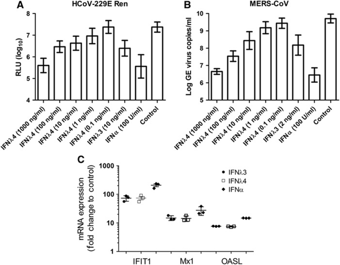
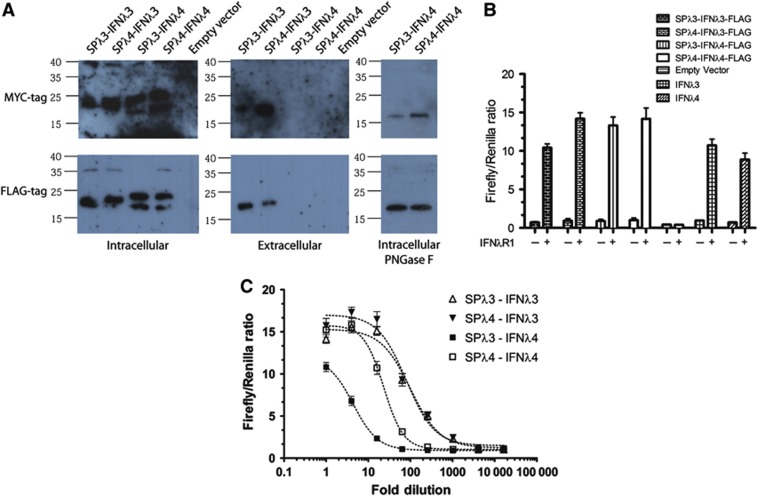
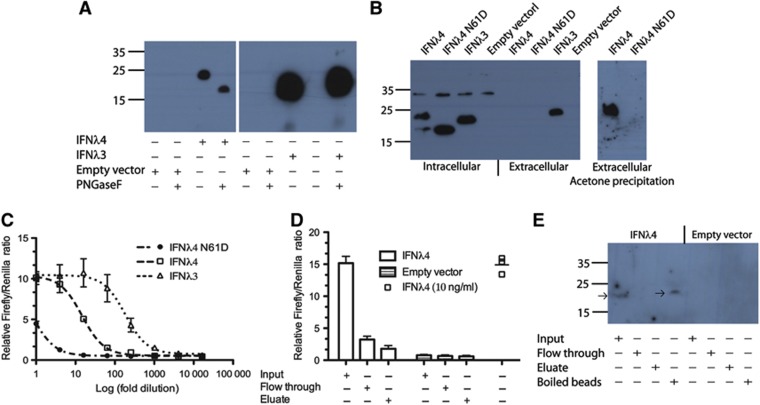
The IFNL4 gene is a recently discovered type III interferon, which in a significant fraction of the human population harbours a frameshift mutation abolishing the IFNλ4 ORF. The expression of IFNλ4 is correlated with both poor spontaneous clearance of hepatitis C virus (HCV) and poor response to treatment with type I interferon. Here, we show that the IFNL4 gene encodes an active type III interferon, named IFNλ4, which signals through the IFNλR1 and IL-10R2 receptor chains. Recombinant IFNλ4 is antiviral against both HCV and coronaviruses at levels comparable to IFNλ3. However, the secretion of IFNλ4 is impaired compared to that of IFNλ3, and this impairment is not due to a weak signal peptide, which was previously believed. We found that IFNλ4 gets N-linked glycosylated and that this glycosylation is required for secretion. Nevertheless, this glycosylation is not required for activity. Together, these findings result in the paradox that IFNλ4 is strongly antiviral but a disadvantage during HCV infection.


Conflict of interest statement

The authors declare that they have no conflict of interest.

Figures

Figure 1
Figure 1
Purification of recombinant human interferon lambda 4. (A) Refolded IFNλ4 was loaded on an anion exchange column (5 ml Hi trap SP FF). The protein was eluted from the column using a salt gradient and the fractions of the peak at ≈10 ml were collected. (B) The fractions collected from the ion exchange column were applied to a gelfiltration column (Hi load Superdex 75). The fractions around the peak at≈75 ml were pooled. (C) A Coomassie stained 12% SDS–PAGE gel of the purified protein.
Figure 2
Figure 2
Activity of recombinant human interferon lambda 4. (A) The activity of IFNλ4 was tested and compared to IFNλ3 in HL-116 reporter cells. Dose response of IFNλ3 and IFNλ4 was performed in triplicate. Mean and s.d. are shown. (B) HepG2 cells were treated with IFNα (1000 U/ml), IFNλ3 (10 ng/ml) or IFNλ4 (10 ng/ml). After 4 h, the level of the interferon-induced genes, IFIT1, MX1 and OASL, was quantified by qPCR, four independent experiments are shown, mean and s.e.m. are plotted. (C) HEK293 cells were transfected with the IFNλR1 and/or treated with siRNA against IL-10R2 as indicated. The cells were also transfected with Renilla and Firefly luciferase reporter constructs. The Firefly construct is IFN inducible whereas the Renilla is constitutively expressed. The cells were subsequently either treated with IFNλ3, IFNλ4 or left untreated, treatment with IFNα was performed as a control. (D) HEK293 cells were transfected with IFNλR1 as well as Renilla and Firefly luciferase reporter constructs (as in C). The cells were then treated with anti IL-10R2 antibody or control antibody followed by interferon treatment as indicated.
Figure 3
Figure 3
Homology model of human interferon lambda 4. (A) Alignment of IFNλ3 and IFNλ4 using Clustal W. The position of the signal peptide (SP) in IFNλ3 and IFNλ4 is shown. The positions of the helices in IFNλ3 and in the model of IFNλ4 shown in (B) are shown. A possible N-linked glycosylation site at Asn61 is marked by a square in IFNλ4. (B) Homology model of IFNλ4 (homIFNλ4) generated by the Swiss model server using IFNλ3 (PDB entry code HHC3) as a template. The position of the individual structural elements is denoted as AF. The cysteins are shown as yellow sticks. The positions of C76 and C178 that could form a disulphide bridge specific for IFNλ4 are shown. (C) Superimposition of IFNλ3 (blue) and homIFNλ4 (red). (D) Comparison of helix D of IFNλ3 (blue) and homIFNλ4 (red). The residues that are expected to interact with IL-10R2 are labelled in blue for IFNλ3 and red for IFNλ4. Disulphides are shown in yellow. (E) Superimposition of homIFNλ4 (red) onto IFNλ1 (green) bound to IFNλR1 (cyan). N61 of homIFNλ4 (red) and W47 of IFNλ1 (green) are shown in sticks and labelled.
Figure 4
Figure 4
Antiviral effect of recombinant human Interferon lambda 4 against hepatitis C virus. (A, B) The antiviral effect of IFNα (A), IFNλ3 or IFNλ4 (B) against replication of the HCV JcR-2a chimaera in the Huh7-Lunet N hCD81-FLuc cell line is shown. (C, D) The antiviral effect of IFNα (C), IFNλ3 or IFNλ4 (D) against replication of the HCV JcR-2a chimaera in the HepG2 cell line is shown. The plot shows the average of three independent experiments with the Renilla luciferase activity normalised to the untreated control. Background luciferase activity is 0.2% for (A, B) and 2.4% for (C, D).
Figure 5
Figure 5
Antiviral effect of IFNλ4 in human airway epithelial (HAE) cell culture. (A) The antiviral effect of IFNα, IFNλ3 and IFNλ4 against the human coronavirus HCoV-229E expressing luciferase was tested in human HAE cultures. For each data point, triplicate measurements were performed on three different donors, mean and s.d. are shown. (B) The antiviral effect of IFNα, IFNλ3 and IFNλ4 against the coronavirus MERS-CoV was tested in human HAE cultures. As in (A), experiments were performed in triplicate on each of the three donors, mean and s.d. are shown. (C) HAE cell cultures were treated with IFNα (100 U/ml), IFNλ3 (10 ng/ml) or IFNλ4 (10 ng/ml). After 72 h, the level of the interferon-induced genes IFIT1, MX1 and OASL were quantified by qPCR, mean and s.e.m. are indicated.
Figure 6
Figure 6
Secretion of human interferon lambda 4. (A) Constructs of IFNλ3 and IFNλ4 containing either the IFNλ3 or IFNλ4 signal peptide were transfected into HEK293 cells using pcDNA3.1 as a control. Western blots were performed on the intracellular and extracellular fractions as shown, using antibodies against the MYC or FLAG tag. For the cell fraction, the samples of IFNλ4 were also subjected to treatment with PNGase F to confirm the presence of N-linked glycosylation. (B, C) The extracellular supernatants from HEK293 cells transfected with the FLAG-tagged constructs from (A) were added to HEK293 cells transfected with the pEF2 vector containing IFNλR1 as well as the interferon inducible luciferase reporter system (see Figure 2C), in order to measure IFNλ activity present in the extracellular supernatants. In (B), the extracellular fraction was undiluted whereas in (C) a serial dilution was performed.
Figure 7
Figure 7
The secreted glycosylated IFNλ4 is active. (A) Western blot of acetone precipitated media from cells transfected with empty vector or FLAG-tagged IFNλ3 or IFNλ4. Where indicated the sample was subjected to deglycosylation using PNGase F. (B) Western blot of intracellular and extracellular fractions of cells transfected with empty vector or FLAG-tagged IFNλ3, IFNλ4 or the IFNλ4 N61D mutant. (C) HEK293 cells were transfected with IFNλR1 as well as the interferon inducible luciferase reporter system (see Figure 2C). The cells were subsequently treated with the media from (B) and the luciferase activities were measured. (D) Measurement of IFNλ activity was performed as in (C). Input: media before addition of Con A beads, Flow through: flow through from the Con A beads and Eluate: eluate from the Con A beads. (E) Western blot of the samples from (D) as well as the boiled Con A beads. The arrows show the position of IFNλ4.

Similar articles

Cited by

References

    1. Ank N, West H, Bartholdy C, Eriksson K, Thomsen AR, Paludan SR (2006) Lambda interferon (IFN-lambda), a type III IFN, is induced by viruses and IFNs and displays potent antiviral activity against select virus infections in vivo. J Virol 80: 4501–4509 - PMC - PubMed
    1. Bibert S, Roger T, Calandra T, Bochud M, Cerny A, Semmo N, Duong FH, Gerlach T, Malinverni R, Moradpour D, Negro F, Mullhaupt B, Bochud PY (2013) IL28B expression depends on a novel TT/-G polymorphism which improves HCV clearance prediction. J Exp Med 210: 1109–1116 - PMC - PubMed
    1. Bitzegeio J, Bankwitz D, Hueging K, Haid S, Brohm C, Zeisel MB, Herrmann E, Iken M, Ott M, Baumert TF, Pietschmann T (2010) Adaptation of hepatitis C virus to mouse CD81 permits infection of mouse cells in the absence of human entry factors. PLoS Pathog 6: e1000978. - PMC - PubMed
    1. Booth D, George J (2013) Loss of function of the new interferon IFN-lambda4 may confer protection from hepatitis C. Nat Genet 45: 119–120 - PubMed
    1. Bordoli L, Kiefer F, Arnold K, Benkert P, Battey J, Schwede T (2009) Protein structure homology modeling using SWISS-MODEL workspace. Nat Protoc 4: 1–13 - PubMed

Publication types

MeSH terms