Comparative Study
. 2013 Dec;112(9):1775-91.
doi: 10.1093/aob/mct232. Epub 2013 Oct 29.

Floral elaiophores in Lockhartia Hook. (Orchidaceae: Oncidiinae): their distribution, diversity and anatomy

Affiliations
Comparative Study

Floral elaiophores in Lockhartia Hook. (Orchidaceae: Oncidiinae): their distribution, diversity and anatomy

Mario A Blanco et al. Ann Bot. 2013 Dec.

Abstract

Background and aims: A significant proportion of orchid species assigned to subtribe Oncidiinae produce floral oil as a food reward that attracts specialized bee pollinators. This oil is produced either by glabrous glands (epithelial elaiophores) or by tufts of secretory hairs (trichomal elaiophores). Although the structure of epithelial elaiophores in the Oncidiinae has been well documented, trichomal elaiophores are less common and have not received as much attention. Only trichomal elaiophores occur in the genus Lockhartia, and their distribution and structure are surveyed here for the first time.

Methods: Flowers of 16 species of Lockhartia were studied. The location of floral elaiophores was determined histochemically and their anatomical organization and mode of oil secretion was investigated by means of light microscopy, scanning electron microscopy and transmission electron microscopy.

Key results and conclusions: All species of Lockhartia investigated have trichomal elaiophores on the adaxial surface of the labellum. Histochemical tests revealed the presence of lipoidal substances within the labellar trichomes. However, the degree of oil production and the distribution of trichomes differed between the three major groups of species found within the genus. All trichomes were unicellular and, in some species, of two distinct sizes, the larger being either capitate or apically branched. The trichomal cuticle was lamellate, and often appeared distended due to the subcuticular accumulation of oil. The labellar trichomes of the three species examined using transmission electron microscopy contained dense, intensely staining cytoplasm with apically located vacuoles. Oil-laden secretory vesicles fused with the plasmalemma and discharged their contents. Oil eventually accumulated between the cell wall and cuticle of the trichome and contained electron-transparent profiles or droplets. This condition is considered unique to Lockhartia among those species of elaiophore-bearing Oncidiinae studied to date.

Keywords: Anatomy; Lockhartia; Oncidiinae; Orchidaceae; callus; elaiophore; oil secretion; trichomes.


Figures

Fig. 1.
Fig. 1.
Variation in floral morphology in the genus Lockhartia, illustrated by species included in the present study. (A) L. acuta (Blanco 3221). (B) L. grandibractea (Blanco 2559). (C) L. lepticaula (Blanco 3237). (D) L. lunifera (Blanco 2688). (E) L. micrantha (Blanco 2562). (F) L. oerstedii (Blanco 2565). (G) L. hercodonta (Blanco 3232); flower shown upright, but in this species the flowers are usually pendent. (H) L. obtusata (Blanco 3025). (I) L. bennettii (Whitten 1704, photograph courtesy of W. M. Whitten). Scale bars = 5 mm.
Fig. 2.
Fig. 2.
Labellar trichomes of Lockhartia species from the Imbricata group prior to (left) and following (right) staining with Sudan dyes. (A, B) L. micrantha (Blanco 2562). (C, D) L. lunifera (Blanco 2688). (E, F) L. verrucosa (Blanco 3230). (G, H) L. acuta (Blanco 3221). In (A–F) the trichomes are located in a shallow concavity on the proximal part of the callus (i.e. the elaiophore cushion; see text for details). Scale bars = 1 mm.
Fig. 3.
Fig. 3.
Labellar trichomes of Lockhartia species from the Longifolia group prior to (left) and following (right) staining with Sudan dyes. (A, B) L. hercodonta. (C, D) L. longifolia. Scale bars = 1 mm.
Fig. 4.
Fig. 4.
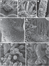
Labellar trichomes of (A–C) Lockhartia lunifera and (D–G) L. oerstedii viewed by scanning electron microscopy. (A) Long trichomes at base of labellum. (B) Surface of trichomes with striate and blistered cuticle, and residues of secreted material visible at apex (asterisk). (C) Detail of middle portion of trichome with secretion (asterisk). (D) Long trichomes reclining on labellum surface and surrounded by shorter, erect trichomes. Stomata (arrows) occur in the epidermis directly behind the secretory area. (E) Long trichomes with branched tips. (F) Branched apices of long trichomes. (G) Long trichomes with striate and blistered cuticle towards apex (arrows indicate cuticle blisters). Scale bars (A, D, E) = 200 μm; (B, C, F, G) = 20 μm.
Fig. 5.
Fig. 5.
Labellar trichomes of Lockhartia verrucosa viewed by scanning electron microscopy. (A) Elaiophore at base of labellum; base of callus (ca) and column (co) visible. (B) Detail of elaiophore; notice long trichomes surrounded by short trichomes. (C) Branched and rounded apices of long and short trichomes, respectively. (D) Cuticular blisters (arrows) at apex of long trichomes. Scale bars (A, B) = 400 μm; (C, D) = 100 μm.
Fig. 6.
Fig. 6.
Labellar trichomes of (A–D) Lockhartia acuta, (E, F) L. bennettii and (G–I) L. obtusata viewed by scanning electron microscopy. In A–C and G, the base (proximal part) of the labellum is towards the top of the image. (A) Callus clothed with short trichomes; note the tuft of longer, capitate trichomes at base of labellum. (B) Tuft of capitate trichomes at base of labellum. (C) Detail of apical portion of capitate trichomes. (D) Detail of short trichomes from main part of the callus. (E) Part of the callus, epidermis obscured by trichomes. (F) Apical portion of trichome with secretory residues. (G) Callus excised from the rest of the labellum, showing basal trapezoid projection, semi-circular rim and apical tooth, all of which are partly clothed with trichomes. (H) Group of trichomes on rim surface. (I) Apex of trichome, showing unidentified crystalline deposits. Scale bars (A, E, G) = 750 μm; (B, D, H) = 100 μm; (C, F, I) = 25 μm.
Fig. 7.
Fig. 7.
Labellar trichomes of Lockhartia oblongicallosa viewed by scanning electron microscopy. The proximal part (base) of the labellum is towards the top of the images. (A) Base of labellum showing two parallel keels and intervening area partly clothed with trichomes. (B) Trichomes at base of callus. (C) Detail of capitate trichomes near the central region of callus. Scale bars = 500 μm.
Fig. 8.
Fig. 8.
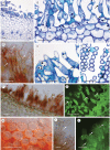
Transverse sections of the trichome-bearing region of the labellum (i.e. the elaiophore) of (A, B) Lockhartia niesseniae, (C) L. acuta, (D) L. bennettii, (E, F) L. oerstedii and (G) L. verrucosa, and secretory trichomes of L. verrucosa (H, I) viewed by light microscopy. (A) Base of labellum of L. niesseniae showing elaiophore trichomes, subepidermal parenchyma and ground parenchyma with vascular bundles. The concavity that contains the trichomes is part of the ‘elaiophore cushion’. (B) Detail of (A): secretory trichomes and adaxial epidermal cells. (C) Base of labellum of L. acuta: section through the group of long trichomes. In (A–C) the long elaiophore trichomes lie parallel to the longitudinal axis of the labellum and are shown in transverse section; the short elaiophore trichomes are shown in oblique section. (D) Central part of labellum of L. bennettii showing adaxial epidermal cells, many of which are trichomatous; the trichomes mostly lean towards the labellum apex and are cut obliquely. (E) Base of labellum of L. oerstedii showing sections through long and short trichomes. Idioblasts with phenolic contents and raphides (arrows) occur in the ground parenchyma. (F) Detail of E, showing trichomatous epidermis and subepidermal parenchyma composed of small, densely packed cells. (G) Base of labellum of L. verrucosa with trichomatous adaxial epidermis and subepidermal parenchyma, both tissues intensely stained with TBO. An idioblast with raphides is indicated by the arrow. (H) Lipid droplets on the surface of branched apex of long trichomes (arrows), stained with Sudan III. (I) Intracellular lipids following staining of trichomes with auramine O (arrows). Scale bars = 100 μm.
Fig. 9.
Fig. 9.
Secretory trichomes of Lockhartia lunifera viewed by light microscopy. (A) Transverse section of labellum base stained with TBO and showing trichomatous epidermis, subepidermal parenchyma and ground parenchyma with vascular bundles. Note long elaiophore trichomes cut transversely above adaxial epidermis. (B) Short elaiophore trichomes with dense cytoplasm and nuclei, their surface coated with heterogeneous secretion (arrows). (C) Long trichomes with surface lipids stained with Sudan III (arrows). (D) Heterogeneous secretion (arrows) coating surface of short trichomes. (E) Transverse section of long trichomes with thick cellulose cell wall coated with secretion (arrow). (F) Hand-cut section of labellum stained with Sudan III showing trichomatous epidermis and plastids in subepidermal parenchyma. Lipids are present in trichomes (arrow) and subepidermal parenchyma. (G) Trichomes surrounded by heterogeneous secretion (arrows) following staining with auramine O. (H) Secretory, long trichomes in transverse section with thick cell walls coated with secretion (arrow) that stains with Sudan III. (I) Apex of trichome stained with Sudan III showing blistered cuticle and subcuticular secretion (arrows). (J) Blistered cuticle and secretion stained with auramine O (arrows). Abbreviations: se, secretory epidermis; sp, subepidermal parenchyma. Scale bars = 50 μm.
Fig. 10.
Fig. 10.
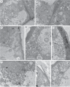
Ultrastructure of secretory cells of (A–E) Lockhartia oerstedii and (F–H) L. lunifera viewed by transmission electron microscopy. (A) Atrichomatous, adaxial epidermal cell showing centrally positioned nucleus, plastids with starch grains, and vacuole towards the bottom of the image. (B) Detail of granular cytoplasm with plastids, mitochondria and endoplasmic reticulum. The cell wall contains primary pit-fields with plasmodesmata. (C) Parietal cytoplasm containing numerous lipid droplets and plastid with starch grain. (D) Secretory vesicles adjacent to cell wall, and vacuoles with myelin-like figures. (E) Secretory vesicles near plasmalemma. Note lamellate structure of the cuticle overlying the wall. (F) Numerous lipid droplets in perinuclear cytoplasm. (G) Lipid droplets in parietal cytoplasm towards base of trichome. (H) Plastids containing starch grains and lipid droplets. Abbreviations: cw, cell wall; d, dictyosome; ER, endoplasmic reticulum; l, lipid-filled vesicle; m, mitochondrion; mf, myelin-like figure; n, nucleus; p, plastid; st, starch; sv, secretory vesicle; v, vacuole. Scale bars = 2 μm.
Fig. 11.
Fig. 11.
Ultrastructure of secretory hairs of Lockhartia verrucosa viewed by transmission electron microscopy. (A) Large lipid droplets and endoplasmic reticulum profiles in parietal cytoplasm. (B) Lipid droplets and numerous small vacuoles. (C) Plastids in parietal cytoplasm containing starch and small lipid droplets. (D) Mitochondria, plastids and small vacuoles in basal part of trichome. (E) Secretory vesicles, mitochondria and dictyosomes adjacent to plasmalemma. Abbreviations: ER, endoplasmic reticulum; l, lipid-filled vesicle; m, mitochondrion; st, starch; sv, secretory vesicle; v, vacuole. Scale bars = 2 μm.

Similar articles

Cited by

References

    1. Aliscioni SS, Torretta JP, Bello ME, Galati BG. Elaiophores in Gomesa bifolia (Sims) M.W. Chase & N.H. Williams (Oncidiinae: Cymbidieae: Orchidaceae): structure and oil secretion. Annals of Botany. 2009;104:1141–1149. - PMC - PubMed
    1. Alves dos Santos I, Machado IC, Gaglianone MC. História natural das abelhas coletoras de óleo. Oecologia Brasiliensis. 2007;11:544–557.
    1. Brummitt RK, Powell CE. Authors of plant names. Kew, UK: Royal Botanical Gardens, Kew; 1992.
    1. Buchman SL. The ecology of oil flowers and their bees. Annual Review of Ecology and Systematics. 1987;18:343–369.
    1. Chase MW. A reappraisal of the oncidioid orchids. Systematic Botany. 1986;11:477–491.

Publication types