. 2014 Jan;13(1):184-203.
doi: 10.1074/mcp.M113.030155. Epub 2013 Oct 29.

Elucidating novel hepatitis C virus-host interactions using combined mass spectrometry and functional genomics approaches

Affiliations

Elucidating novel hepatitis C virus-host interactions using combined mass spectrometry and functional genomics approaches

Marie-Anne Germain et al. Mol Cell Proteomics. 2014 Jan.

Abstract

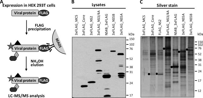
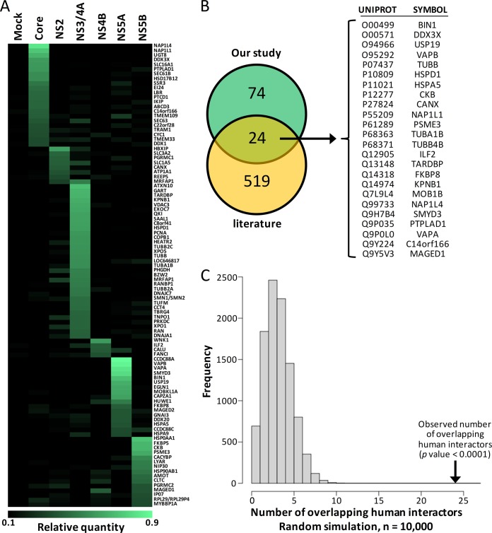
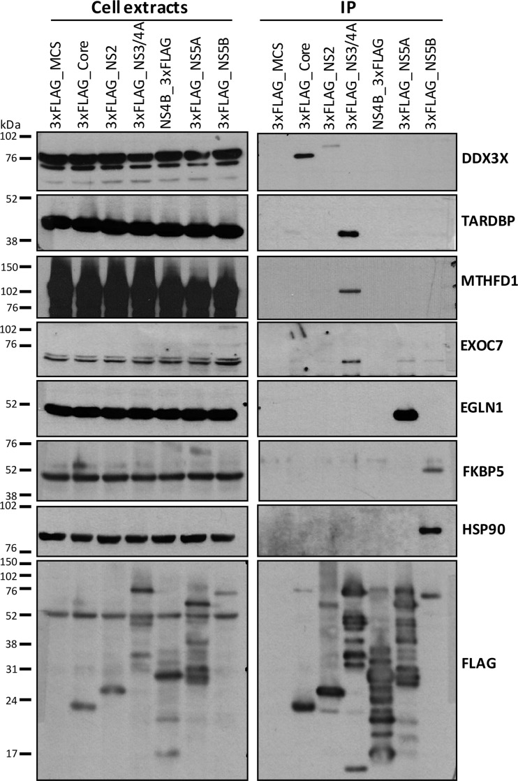
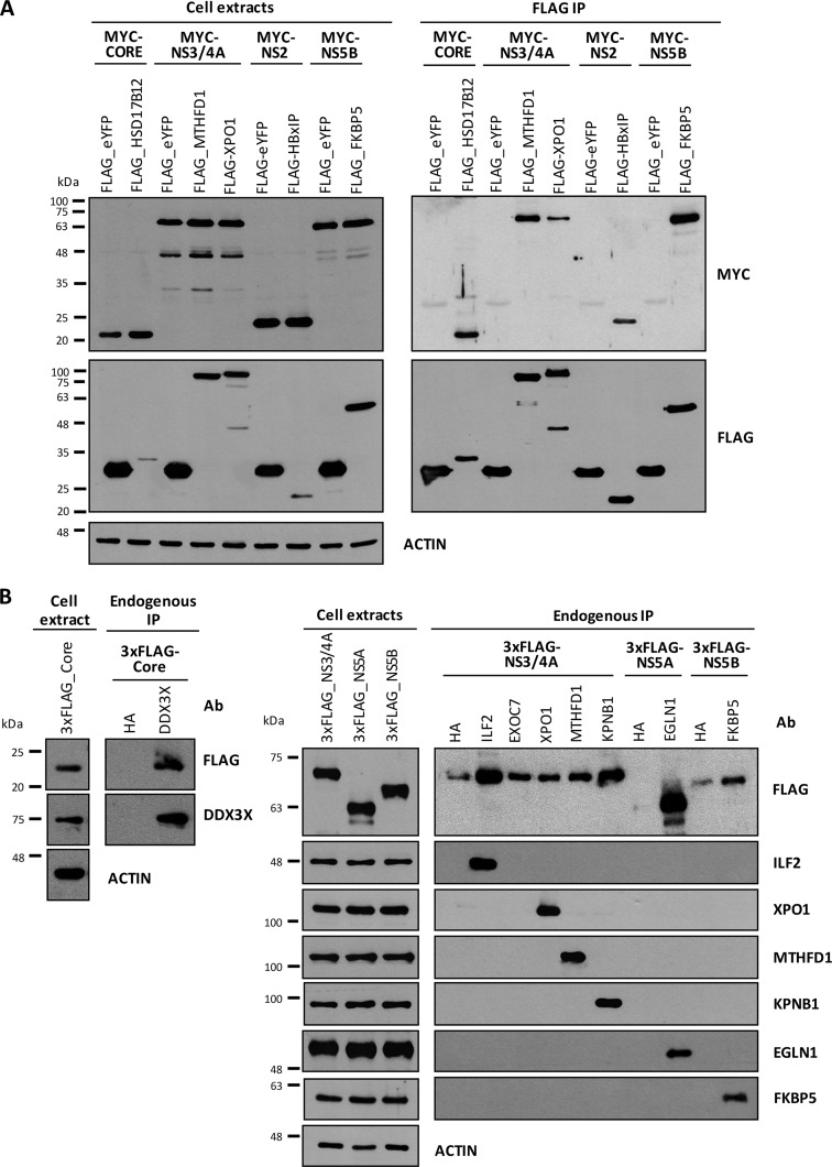
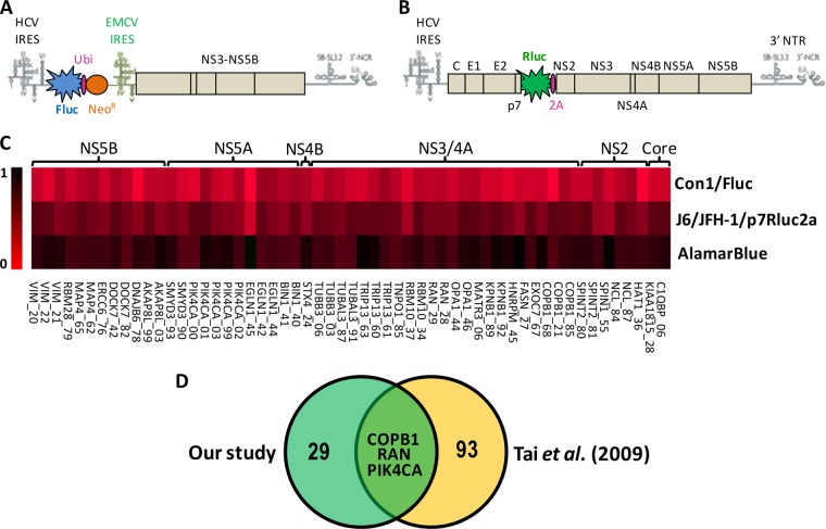
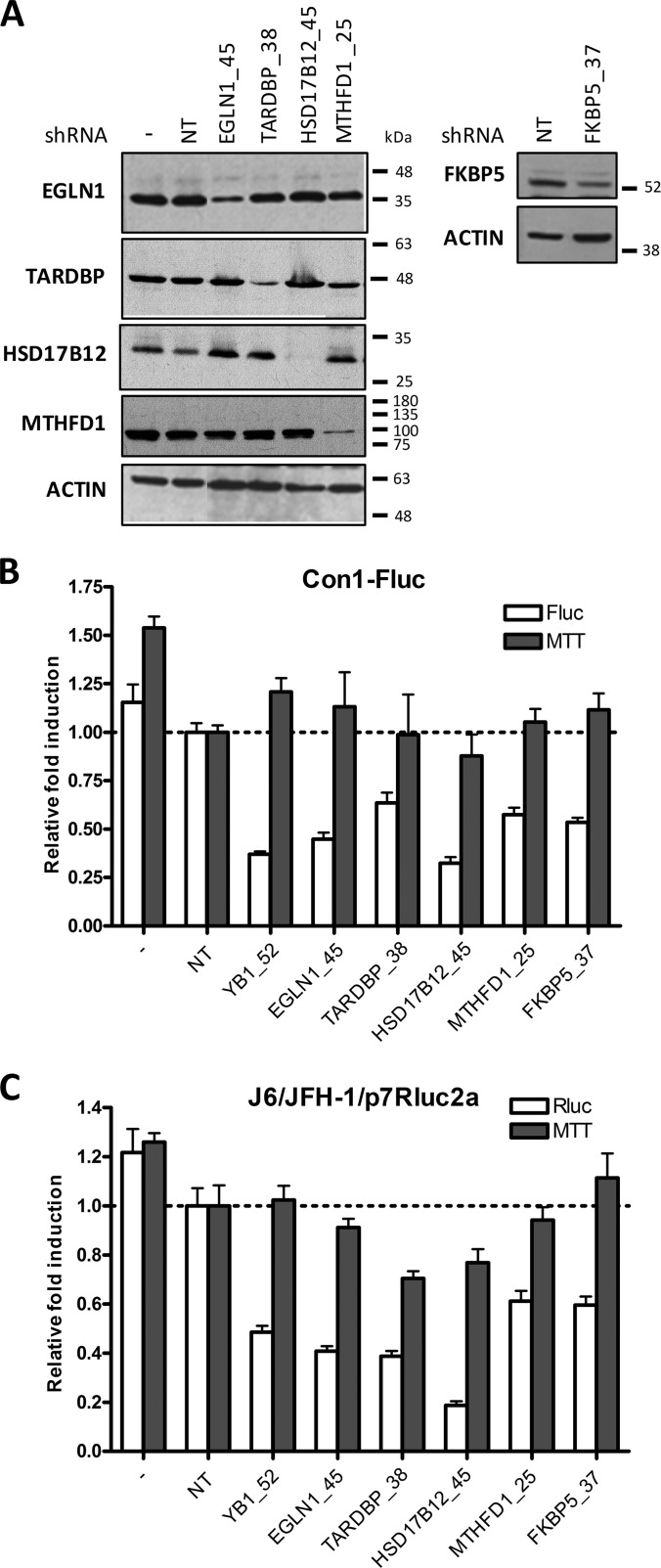
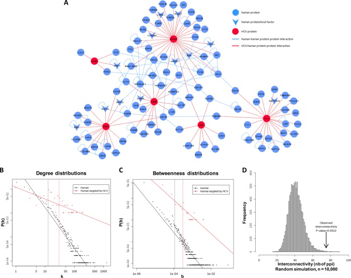
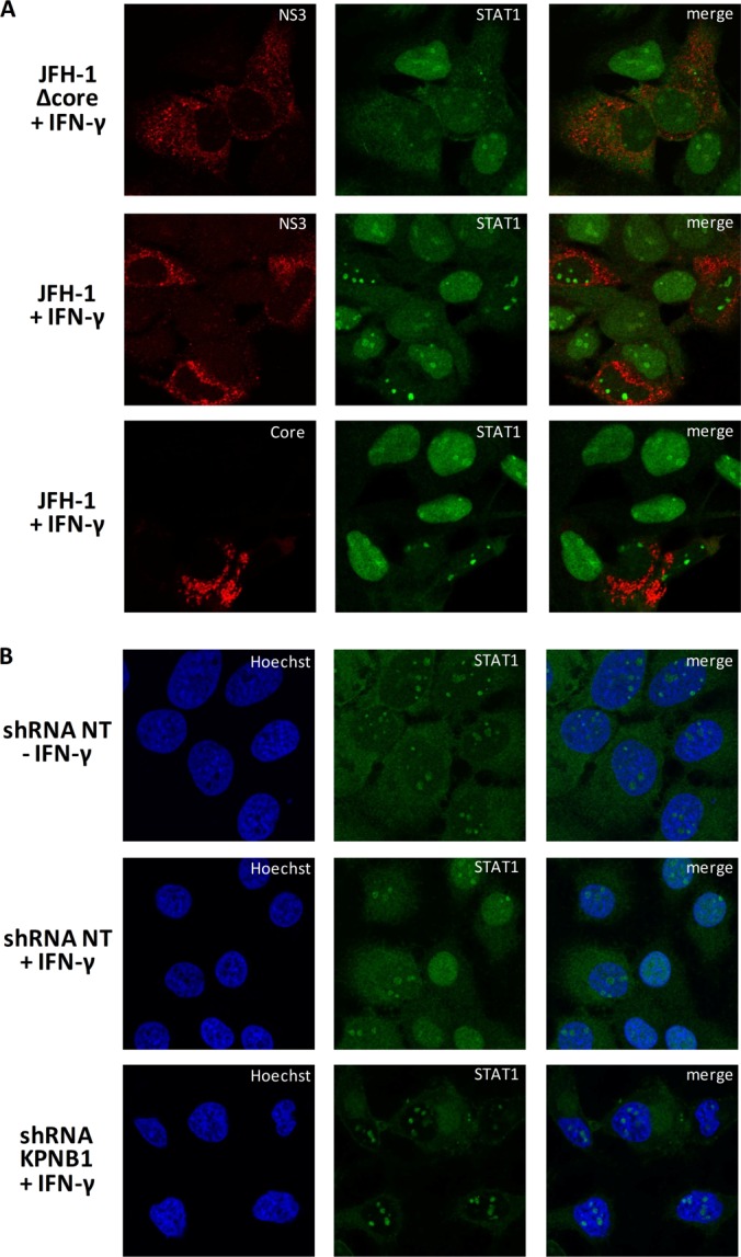
More than 170 million people worldwide are infected with the hepatitis C virus (HCV), for which future therapies are expected to rely upon a combination of oral antivirals. For a rapidly evolving virus like HCV, host-targeting antivirals are an attractive option. To decipher the role of novel HCV-host interactions, we used a proteomics approach combining immunoprecipitation of viral-host protein complexes coupled to mass spectrometry identification and functional genomics RNA interference screening of HCV partners. Here, we report the proteomics analyses of protein complexes associated with Core, NS2, NS3/4A, NS4B, NS5A, and NS5B proteins. We identified a stringent set of 98 human proteins interacting specifically with one of the viral proteins. The overlap with previous virus-host interaction studies demonstrates 24.5% shared HCV interactors overall (24/98), illustrating the reliability of the approach. The identified human proteins show enriched Gene Ontology terms associated with the endoplasmic reticulum, transport proteins with a major contribution of NS3/4A interactors, and transmembrane proteins for Core interactors. The interaction network emphasizes a high degree distribution, a high betweenness distribution, and high interconnectivity of targeted human proteins, in agreement with previous virus-host interactome studies. The set of HCV interactors also shows extensive enrichment for known targets of other viruses. The combined proteomic and gene silencing study revealed strong enrichment in modulators of HCV RNA replication, with the identification of 11 novel cofactors among our set of specific HCV partners. Finally, we report a novel immune evasion mechanism of NS3/4A protein based on its ability to affect nucleocytoplasmic transport of type I interferon-mediated signal transducer and activator of transcription 1 nuclear translocation. The study revealed highly stringent association between HCV interactors and their functional contribution to the viral replication cycle and pathogenesis.


Conflict of interest statement

Conflict of interest: D.L. is a shareholder in ViroCura Therapeutics Inc.

Figures

Fig. 1.
Fig. 1.
Immunopurification of viral protein complexes. A, schematic representation of 3xFLAG-tagged viral protein immunoprecipitation. Briefly, 3xFLAG-tagged viral proteins were expressed in 293T cells and cell lysates were incubated with FLAG resin, extensively washed, and eluted with ammonium hydroxide to enrich viral protein and its attached host–protein complexes before being submitted for LC-MS/MS analysis. B, immunoblot detection of the viral-tag protein at expected weight in cell extracts. C, silver staining of SDS-PAGE gel for the different IP conditions with viral proteins (black dots) and other bands corresponding to co-IP host proteins.
Fig. 2.
Fig. 2.
Heat map representation of enriched HCV-associated host proteins. A, proportions for each of the seven experimental conditions are represented for host protein hits (Σ 7 conditions = 1 for each host protein). The darker color correlates with the absence of the host protein in the condition, and brighter green indicates a high prevalence of the host protein in the condition. Orders of hit representation are according to function of viral protein specificity and are sorted by ascending proportion. B, Venn diagram of the overlap between HCV interactors identified in our study and those reported in the literature with the list of the 24 common interactors. C, statistical analysis of the overlap. The 10,000 overlap sizes obtained via random simulation are plotted. The observed value of x is indicated by the vertical arrow, and its significance is given.
Fig. 3.
Fig. 3.
Validation by immunoprecipitation–Western blotting of selected virus–host protein partners identified by MS analysis. Selected hits retrieved by IP-MS/MS were validated by FLAG IP coupled to FLAG peptide elution and Western blot detection. DDX3X was present and specific to the Core condition; TARDBP, MTHFD1, and EXOC7 were NS3/4A partners; EGLN1 was a specific NS5A interactor; and FKBP5 and HSP90 interacted specifically with NS5B. Flag immunoblots show the expression of viral protein in the lysates and IP conditions.
Fig. 4.
Fig. 4.
Immunoprecipitation of host interactor proteins validates their interactions with viral HCV proteins. A, immunoprecipitation of FLAG-tagged host interactor proteins HSD17B12, MTHFD1, XPO1, HBxIP, and FKBP5 coupled to FLAG peptide elution and Western blotting detection of Myc-tagged HCV proteins (reciprocal co-IP). Immunoprecipitation of FLAG-eYFP was used as a negative control. B, immunoprecipitation of endogenous host interactor proteins DDX3X, ILF2, EXOC7, XPO1, MTHFD1, KPNB1, EGLN1, and FKBP5 followed by Western blotting detection of 3xFLAG-tagged HCV proteins (reciprocal co-IP). Immunoprecipitation of Human influenza hemagglutinin (HA) was used as a negative control. Ab, antibody.
Fig. 5.
Fig. 5.
Effect of silencing potential HCV-associated host proteins by shRNA on HCV viral replication. A, B, schematic representation of the HCV reporter systems used in the shRNA screen. A, Huh7.5-Con1-Fluc subgenomic replicon contains the 5′UTR and 3′UTR of the HCV RNA genome that are required for its replication and expresses Fluc under the control of the HCV internal ribosome entry site and the viral replicative unit (protein NS3 to NS5B of genotype 1b) under an encephalomyocarditis virus internal ribosome entry site. B, J6/JFH-1/p7Rluc2a chimeric construct is composed of the HCV 5′UTR region extending to NS2 from a J6 sequence, genetically engineered to express Rluc, and the sequence from NS3 through the 3′UTR from the JFH-1 isolate (genotype 2a). The reporter protein is inserted between p7 and NS2 with a foot-and-mouth-disease virus peptide cleavage site resulting in self-cleavage from the polyprotein. C, TIGR MultiExperiment Viewer (TMEV) representation of the shRNA screen results for 32 gene hits whose silencing significantly inhibited HCV viral replication with no observed changes in cell viability. Results are presented as an inhibition heat map and were normalized according to cells treated with shRNA NT (negative control set to 1 (black)) based on an average of three independent experiments. The following criteria were applied to select hits: >25% decrease of viral replication on J6/JFH-1 model; AlamarBlue effect of <25%; and >50% decrease in Con1b system with two shRNAs or >70% if only one shRNA was efficient. Hits are clustered by their corresponding viral binding partners, and the last two digits of the hit name correspond to the shRNA TRC number from the Sigma bank. D, comparison of host factors identified in this study and those from a genome-wide screen by Tai et al. (7).
Fig. 6.
Fig. 6.
Validation of the silencing efficiency of selected shRNA targeting HCV cofactors and their effect on viral replication. A, the gene silencing effect was evaluated for each cofactor via Western blot detection. A decrease of the target protein was observed with the gene-specific shRNA, illustrating the efficacy of gene silencing. Actin immunoblot was performed as a loading control. B, C, the gene silencing effects of shRNA-expressing lentiviruses for each cofactor were determined on HCV replication-dependent reporter activities (MOI of 10) at day 3 post-transduction with HCV replicon-containing cells (B) and in the infection assay (C). The cellular viability was assessed with a 3-(4,5-dimethylthiazol-2-yl)-2,5-diphenyltetrazolium bromide cytotoxicity assay performed in parallel.
Fig. 7.
Fig. 7.
Interaction network of host interactors with viral proteins and degree, betweenness, and interconnectivity of 98 interactors. A, HCV–human protein–protein interaction network from this work. Viral proteins were manually connected to their hit host proteins and host proteins were connected together automatically using Cytoscape. Red nodes: HCV protein. Blue nodes: human protein. Red lines: HCV–human protein–protein interactions identified in this work. Blue lines: human–human protein–protein interactions. Arrowhead nodes: cofactors identified in this work. Nodes are disposed according to a force-directed layout. B, degree distributions of human proteins and human proteins targeted by HCV proteins in the human interactome. P(k) is the probability that a node will connect to k other nodes in the network. Solid lines represent linear regression fits. Vertical dashed lines indicate the mean degree of each distribution. C, betweenness distributions of human proteins and human proteins targeted by HCV proteins in the human interactome. P(b) is the probability that a node will have a betweenness value of b in the network. Solid lines represent linear regression fits. Vertical dashed lines indicate the mean betweenness value for each distribution. D, statistical analysis of the overlap size. The 10,000 overlap sizes obtained via random simulation are plotted. The observed value of x is indicated by the vertical arrow, and its significance is given.
Fig. 8.
Fig. 8.
NS3/4A protein disrupts the type I IFN response. A, an ISRE luciferase reporter assay was performed to examine the effect of viral proteins on the response to type I IFN. HeLa cells were transfected with plasmids encoding viral proteins (NS2, NS3, NS3/4A, or serine protease inactive NS3/4A S139A mutant) and reporter plasmids (pISRE-Fluc and pRL-SV40 Rluc, Stratagene). 48 h post-transfection, cells were treated with 800 IU/ml IFN-β for 6 h, and then luciferase activity was determined using Dual-Glo Luciferase (Promega). Data are expressed as the percentage of luciferase activity relative to the signal obtained with HCV NS2. B, most representative subcellular localizations of STAT1, NS3, and NS3/4A. The effect of HCV NS3/4A on STAT1 subcellular localization was examined via immunofluorescence assay. HeLa cells were co-transfected with plasmids encoding 3xFLAG-NS3 or -NS3/4A and myc-STAT1. 48 h post-transfection, cells were treated with 1,000 IU/ml IFN-β for 45 min. Cells were fixed, permeabilized, stained with anti-FLAG (red) or anti-myc (green) antibody, and examined with a laser-scanning confocal microscope. C, relative localization of STAT1 in nucleus, cytoplasm, or both was analyzed in 100 cells expressing NS3 or NS3/4A. D, effect of NS3 protease activity on STAT1 phosphorylation. HeLa cells were transfected with plasmids encoding indicated viral proteins with or without myc-STAT1 plasmid. 48 h post-transfection, cells were either untreated or treated with 1,000 IU/ml IFN-β for 30 min and lysed. STAT1 tyrosine phosphorylation was assessed by immunoblotting of total lysates with anti-pY701 STAT1 and anti-STAT1 antibodies. Viral protein expression was detected with anti-FLAG antibody.
Fig. 9.
Fig. 9.
NS3/4A and its interactor KPNB1 prevent STAT1 nuclear localization. A, Huh7.5 cells were transfected with JFH-1 mutant Δcore 153–167 or NS3+ Q221L. Six days post-transfection, cells were serum starved for 4 h and then stimulated with 5 ng/ml of IFN-γ for 30 min. Cells were fixed, permeabilized, and stained with anti-NS3 or anti-core (red) and anti-STAT1 (green) and then examined via laser-scanning microscopy. B, Huh7 cells were infected with lentiviruses encoding shRNA NT or KPNB1 for three days (MOI = 10). Three days post-infection, cells were serum starved for 4 h and then stimulated with 5 ng/ml of IFN-γ for 30 min. Cells were fixed, permeabilized, and stained with anti-STAT1 (green). Nuclei were stained with Hoechst. Cells were examined via laser-scanning confocal microscopy.

Similar articles

Cited by

References

    1. Poynard T., Yuen M. F., Ratziu V., Lai C. L. (2003) Viral hepatitis C. Lancet 362, 2095–2100 - PubMed
    1. Welsch C., Jesudian A., Zeuzem S., Jacobson I. (2012) New direct-acting antiviral agents for the treatment of hepatitis C virus infection and perspectives. Gut 61 Suppl 1, i36–i46 - PubMed
    1. Blanchard E., Belouzard S., Goueslain L., Wakita T., Dubuisson J., Wychowski C., Rouille Y. (2006) Hepatitis C virus entry depends on clathrin-mediated endocytosis. J. Virol. 80, 6964–6972 - PMC - PubMed
    1. Tsukiyama-Kohara K., Iizuka N., Kohara M., Nomoto A. (1992) Internal ribosome entry site within hepatitis C virus RNA. J. Virol. 66, 1476–1483 - PMC - PubMed
    1. Romero-Brey I., Merz A., Chiramel A., Lee J. Y., Chlanda P., Haselman U., Santarella-Mellwig R., Habermann A., Hoppe S., Kallis S., Walther P., Antony C., Krijnse-Locker J., Bartenschlager R. (2012) Three-dimensional architecture and biogenesis of membrane structures associated with hepatitis C virus replication. PLoS Pathog. 8, e1003056. - PMC - PubMed

Publication types