

Off-target effects of plasmid-transcribed shRNAs on NFκB signaling pathway and cell survival of human melanoma cells
- PMID: 24170218
- PMCID: PMC3835955
- DOI: 10.1007/s11033-013-2817-7
Off-target effects of plasmid-transcribed shRNAs on NFκB signaling pathway and cell survival of human melanoma cells
Abstract
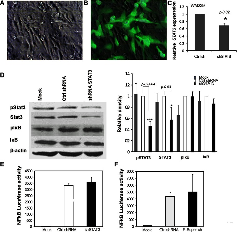
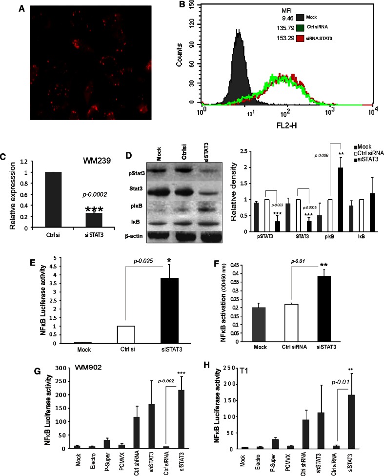
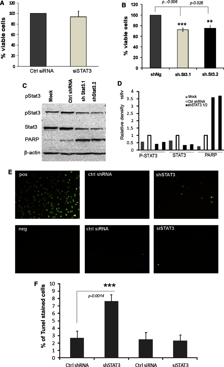
Signal transducer and activator of transcription 3 (STAT3) and nuclear factor kappa-light-chain-enhancer of activated B cells (NFκB) are transcription factors involved in cell survival, inflammation and metastasis. Constitutively activated STAT3 is found in many cancers, including melanoma. To study the crosstalk between STAT3 and NFκB signaling and its role in regulation of cancer cell survival, we used RNA interference (RNAi) to down-regulate STAT3 expression in human melanoma cells. RNAi strategies including double-stranded RNA, small interfering RNA (siRNA), short hairpin RNA (shRNA) and microRNA are widely used to knock down disease-causing genes in a targeted fashion. We found that shRNAs up-regulate non-specific NFκB activity, while siRNA directed against STAT3 specifically increase NFκB activity. The basal survival of melanoma cells is unaffected by STAT3 knockdown-likely due to activation of pro-survival NFκB signaling. Whereas, owing to off-target effects, plasmid-transcribed shRNA affects melanoma survival. Our data show that shRNA-mediated gene silencing induces non-specific or off-target effects that may influence cell functions.
Figures
Similar articles
-
Search for novel STAT3-dependent genes reveals SERPINA3 as a new STAT3 target that regulates invasion of human melanoma cells.Lab Invest. 2019 Nov;99(11):1607-1621. doi: 10.1038/s41374-019-0288-8. Epub 2019 Jul 5. Lab Invest. 2019. PMID: 31278347
-
Functional graphene oxide as a plasmid-based Stat3 siRNA carrier inhibits mouse malignant melanoma growth in vivo.Nanotechnology. 2013 Mar 15;24(10):105102. doi: 10.1088/0957-4484/24/10/105102. Epub 2013 Feb 20. Nanotechnology. 2013. PMID: 23425941
-
IL-32γ inhibits cancer cell growth through inactivation of NF-κB and STAT3 signals.Oncogene. 2011 Jul 28;30(30):3345-59. doi: 10.1038/onc.2011.52. Epub 2011 Mar 21. Oncogene. 2011. PMID: 21423208 Free PMC article.
-
Transcriptome and Proteome Analyses of TNFAIP8 Knockdown Cancer Cells Reveal New Insights into Molecular Determinants of Cell Survival and Tumor Progression.Methods Mol Biol. 2017;1513:83-100. doi: 10.1007/978-1-4939-6539-7_7. Methods Mol Biol. 2017. PMID: 27807832 Review.
-
STAT3 targeting by polyphenols: Novel therapeutic strategy for melanoma.Biofactors. 2017 May 6;43(3):347-370. doi: 10.1002/biof.1345. Epub 2016 Nov 29. Biofactors. 2017. PMID: 27896891 Review.
Cited by
-
Pharmacologic inhibition of MLK3 kinase activity blocks the in vitro migratory capacity of breast cancer cells but has no effect on breast cancer brain metastasis in a mouse xenograft model.PLoS One. 2014 Sep 29;9(9):e108487. doi: 10.1371/journal.pone.0108487. eCollection 2014. PLoS One. 2014. PMID: 25264786 Free PMC article.
-
Discovery of a dicer-independent, cell-type dependent alternate targeting sequence generator: implications in gene silencing & pooled RNAi screens.PLoS One. 2014 Jul 2;9(7):e100676. doi: 10.1371/journal.pone.0100676. eCollection 2014. PLoS One. 2014. PMID: 24987961 Free PMC article.
-
Non-Pigmented Ciliary Epithelium-Derived Extracellular Vesicles Loaded with SMAD7 siRNA Attenuate Wnt Signaling in Trabecular Meshwork Cells In Vitro.Pharmaceuticals (Basel). 2021 Aug 27;14(9):858. doi: 10.3390/ph14090858. Pharmaceuticals (Basel). 2021. PMID: 34577558 Free PMC article.
-
OSM potentiates preintravasation events, increases CTC counts, and promotes breast cancer metastasis to the lung.Breast Cancer Res. 2018 Jun 14;20(1):53. doi: 10.1186/s13058-018-0971-5. Breast Cancer Res. 2018. PMID: 29898744 Free PMC article.
References
-
- Catlett-Falcone R, Landowski TH, Oshiro MM, Turkson J, Levitzki A, Savino R, Ciliberto G, Moscinski L, Fernandez-Luna JL, Nunez G, Dalton WS, Jove R. Constitutive activation of Stat3 signaling confers resistance to apoptosis in human U266 myeloma cells. Immunity. 1999;10(1):105–115. doi: 10.1016/S1074-7613(00)80011-4. - DOI - PubMed
-
- Masuda M, Suzui M, Yasumatu R, Nakashima T, Kuratomi Y, Azuma K, Tomita K, Komiyama S, Weinstein IB. Constitutive activation of signal transducers and activators of transcription 3 correlates with cyclin D1 overexpression and may provide a novel prognostic marker in head and neck squamous cell carcinoma. Cancer Res. 2002;62(12):3351–3355. - PubMed
Publication types
MeSH terms
Substances
LinkOut - more resources
Full Text Sources
Other Literature Sources
Medical
Miscellaneous
