

Corni Fructus Containing Formulation Attenuates Weight Gain in Mice with Diet-Induced Obesity and Regulates Adipogenesis through AMPK
- PMID: 24171041
- PMCID: PMC3792538
- DOI: 10.1155/2013/423741
Corni Fructus Containing Formulation Attenuates Weight Gain in Mice with Diet-Induced Obesity and Regulates Adipogenesis through AMPK
Abstract
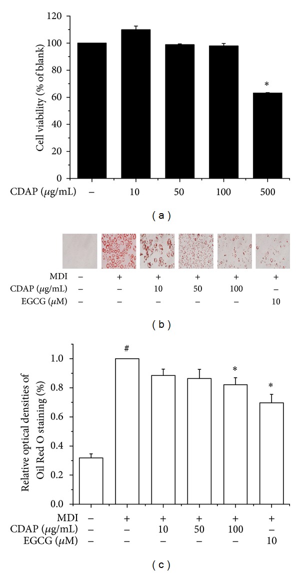
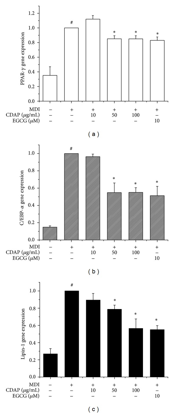
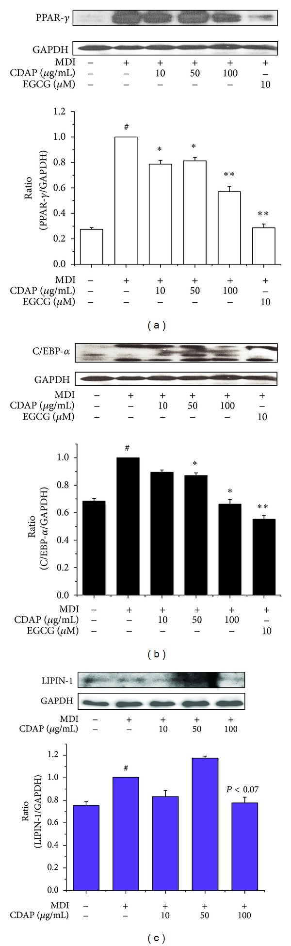
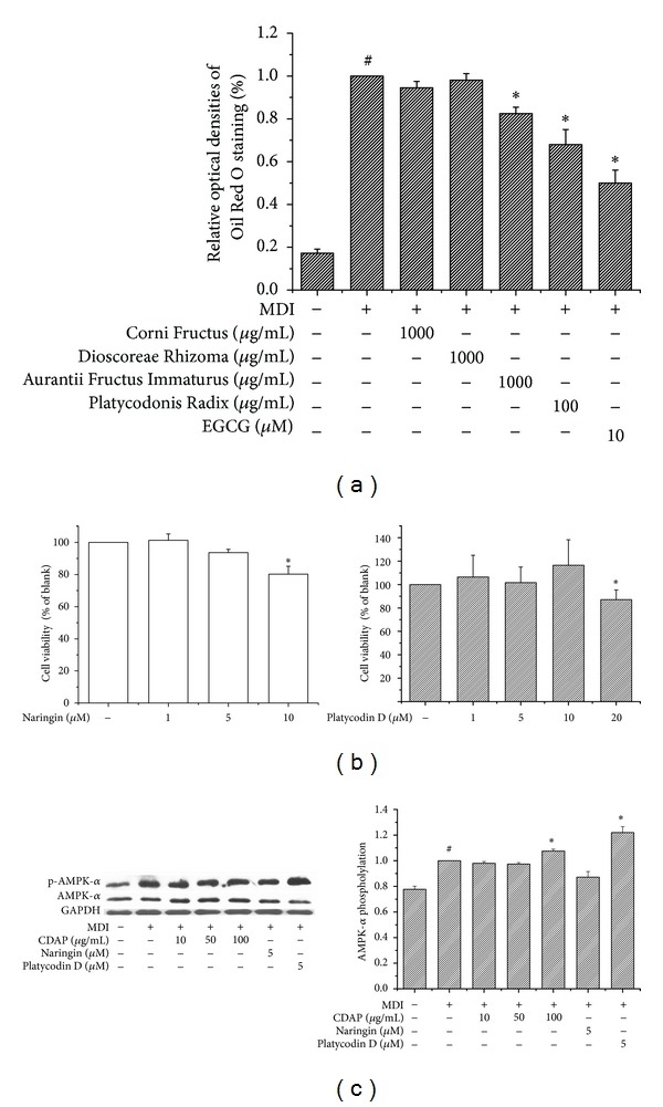
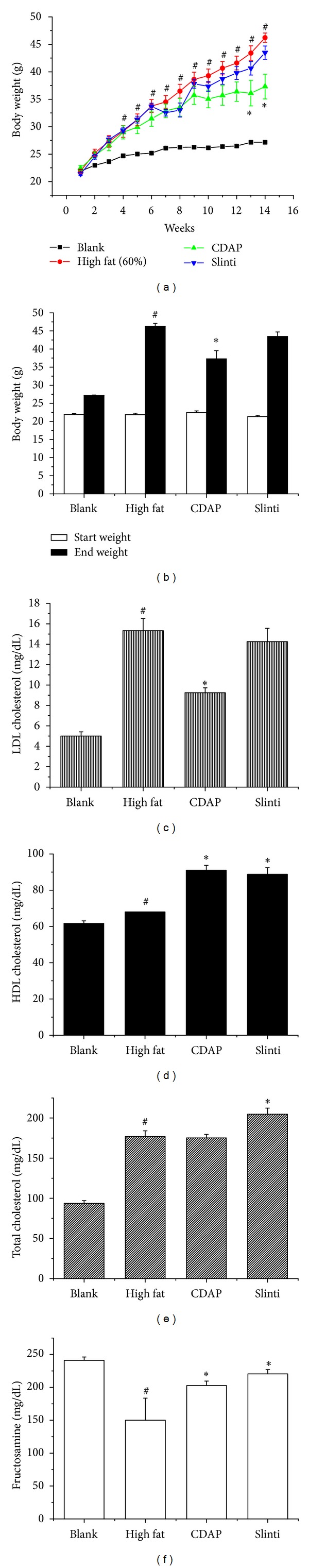
Obesity is a metabolic disorder characterized by chronic inflammation and dyslipidemia and is a strong predictor for the development of hypertension, diabetes mellitus, and cardiovascular disease. This study examined the antiobesity effect of an ethanol extract of Corni Fructus containing formulation (CDAP), which is a combination of four natural components: Corni Fructus, Dioscoreae Rhizoma, Aurantii Fructus Immaturus, and Platycodonis Radix. The cellular lipid content in 3T3-L1 adipocytes was assessed by Oil Red O staining. Expressions of peroxisome proliferator-activated receptor- γ (PPAR- γ ), CCAAT/enhancer-binding protein- α (C/EBP- α ), and lipin-1 were determined by real-time RT-PCR. Western blot was used to determine the protein levels of PPAR- γ , C/EBP- α , and AMP-activated protein kinase- α (AMPK- α ). The CDAP extract suppressed the differentiation of 3T3-L1 adipocytes by downregulating cellular induction of PPAR- γ , C/EBP- α , and lipin-1. The CDAP extract also significantly upregulated phosphorylation of AMPK- α . An in vivo study showed that CDAP induced weight loss in mice with high-fat-diet-induced obesity. These results indicate that CDAP has a potent anti-obesity effect due to the inhibition of adipocyte differentiation and adipogenesis.
Figures
Similar articles
-
Coptis chinensis alkaloids exert anti-adipogenic activity on 3T3-L1 adipocytes by downregulating C/EBP-α and PPAR-γ.Fitoterapia. 2014 Oct;98:199-208. doi: 10.1016/j.fitote.2014.08.006. Epub 2014 Aug 12. Fitoterapia. 2014. PMID: 25128422
-
4-Hydroxyderricin and xanthoangelol from Ashitaba (Angelica keiskei) suppress differentiation of preadiopocytes to adipocytes via AMPK and MAPK pathways.Mol Nutr Food Res. 2013 Oct;57(10):1729-40. doi: 10.1002/mnfr.201300020. Epub 2013 May 16. Mol Nutr Food Res. 2013. PMID: 23681764
-
5,7-Dimethoxyflavone Attenuates Obesity by Inhibiting Adipogenesis in 3T3-L1 Adipocytes and High-Fat Diet-Induced Obese C57BL/6J Mice.J Med Food. 2016 Dec;19(12):1111-1119. doi: 10.1089/jmf.2016.3800. Epub 2016 Nov 9. J Med Food. 2016. PMID: 27828718
-
Polygonum cuspidatum inhibits pancreatic lipase activity and adipogenesis via attenuation of lipid accumulation.BMC Complement Altern Med. 2013 Oct 25;13:282. doi: 10.1186/1472-6882-13-282. BMC Complement Altern Med. 2013. PMID: 24160551 Free PMC article.
-
Beneficial Effect of 7-O-Galloyl-D-sedoheptulose, a Polyphenol Isolated from Corni Fructus, against Diabetes-Induced Alterations in Kidney and Adipose Tissue of Type 2 Diabetic db/db Mice.Evid Based Complement Alternat Med. 2013;2013:736856. doi: 10.1155/2013/736856. Epub 2013 Nov 20. Evid Based Complement Alternat Med. 2013. PMID: 24348717 Free PMC article. Review.
Cited by
-
Veratri Nigri Rhizoma et Radix (Veratrum nigrum L.) and Its Constituent Jervine Prevent Adipogenesis via Activation of the LKB1-AMPKα-ACC Axis In Vivo and In Vitro.Evid Based Complement Alternat Med. 2016;2016:8674397. doi: 10.1155/2016/8674397. Epub 2016 Apr 6. Evid Based Complement Alternat Med. 2016. PMID: 27143989 Free PMC article.
-
Weight loss herbal intervention therapy (W-LHIT) a non-appetite suppressing natural product controls weight and lowers cholesterol and glucose levels in a murine model.BMC Complement Altern Med. 2014 Jul 23;14:261. doi: 10.1186/1472-6882-14-261. BMC Complement Altern Med. 2014. PMID: 25055851 Free PMC article.
-
Effects of Bushen-Jiangya granules on blood pressure and pharmacogenomic evaluation in low-to-medium-risk hypertensive patients: study protocol for a randomized double-blind controlled trial.Trials. 2022 Jan 15;23(1):37. doi: 10.1186/s13063-022-05999-2. Trials. 2022. PMID: 35033168 Free PMC article.
-
Peanut Shell Extract and Luteolin Regulate Lipid Metabolism and Induce Browning in 3T3-L1 Adipocytes.Foods. 2022 Sep 3;11(17):2696. doi: 10.3390/foods11172696. Foods. 2022. PMID: 36076880 Free PMC article.
-
Anti-obesity effects by parasitic nematode (Trichinella spiralis) total lysates.Front Cell Infect Microbiol. 2024 Jan 8;13:1285584. doi: 10.3389/fcimb.2023.1285584. eCollection 2023. Front Cell Infect Microbiol. 2024. PMID: 38259965 Free PMC article.
References
-
- Kang S-I, Ko H-C, Shin H-S, et al. Fucoxanthin exerts differing effects on 3T3-L1 cells according to differentiation stage and inhibits glucose uptake in mature adipocytes. Biochemical and Biophysical Research Communications. 2011;409(4):769–774. - PubMed
-
- Hsieh YH, Wang SY. Lucidone from Lindera erythrocarpa Makino fruits suppresses adipogenesis in 3T3-L1 cells and attenuates obesity and consequent metabolic disorders in high-fat diet C57BL/6 mice. Phytomedicine. 2012;7113(12):420–425. - PubMed
-
- Jessen BA, Stevens GJ. Expression profiling during adipocyte differentiation of 3T3-L1 fibroblasts. Gene. 2002;299(1-2):95–100. - PubMed
-
- Lee H, Bae S, Kim YS, Yoon Y. WNT/β-catenin pathway mediates the anti-adipogenic effect of platycodin D, a natural compound found in Platycodon grandiflorum . Life Sciences. 2011;89(11-12):388–394. - PubMed
LinkOut - more resources
Full Text Sources
Other Literature Sources
