. 2014 Mar 1;23(5):488-501.
doi: 10.1089/scd.2013.0307. Epub 2013 Dec 14.

The role of CCL5 in the ability of adipose tissue-derived mesenchymal stem cells to support repair of ischemic regions

Affiliations

The role of CCL5 in the ability of adipose tissue-derived mesenchymal stem cells to support repair of ischemic regions

Kenichi Kimura et al. Stem Cells Dev. .

Abstract

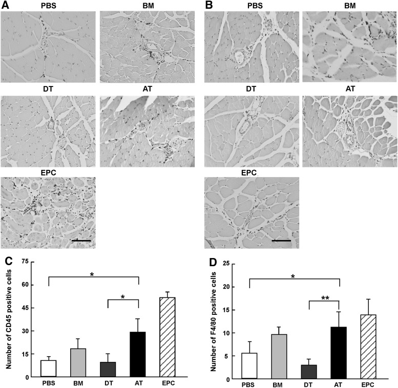
Mesenchymal stem cells (MSC) are multipotent and possess high proliferative activity, and thus are thought to be a reliable cell source for cell therapies. Here, we isolated MSC from adult tissues--bone marrow (BM-MSC), dental tissue (DT-MSC), and adipose tissue (AT-MSC)--to compare how autotransplantation of these MSC effectively supports the repair of bone fracture and ischemic tissue. An analysis by in vitro differentiation assays showed no significant difference among these MSC. The degree of calcification at the joint region of bone fracture was higher in mice transplanted with AT-MSC than in mice transplanted with BM-MSC or DT-MSC. To compare the abilities of MSC, characterize how those MSC affect the repair of ischemic tissue, vascular occlusion was performed by ligation of the femoral artery and vein. Of note, the blood flow in the ischemic region rapidly increased in mice injected with AT-MSC, as contrasted with mice injected with BM- or DT-MSC. The number of CD45- and F4/80-positive cells at the femoral region was higher in AT-MSC recipients than in recipients of BM-MSC or DT-MSC. We evaluated the mRNA expression of angiogenic and migration factors in MSC and found the expression of CCL5 mRNA was higher in AT-MSC than in BM-MSC or DT-MSC. Transplantation of AT-MSC with impaired expression of CCL5 clearly showed a significant delay in the recovery of blood flow compared with the control. These findings have fundamental implications for the modulation of AT-MSC in the repair of vasculature and bone fracture.


Figures

<b>FIG. 1.</b>
FIG. 1.
Isolation of mesenchymal stem cells (MSCs) from human bone marrow (BM), dental tissue (DT), and adipose tissue (AT). (A) BM-, DT-, and AT-MSC showed the spindle-shaped and fibroblast-like morphology. Scale bar indicates 100 μm. (B) Each type of MSC was analyzed to determine osteogenic differentiation potential. Osteogenic differentiation was examined by Alizarin Red S staining without induction (left column) or with induction (right column). There was no difference in BM-, DT-, and AT-MSC. Scale bar indicates 100 μm. (C) Adipogenic differentiation was analyzed by the detection of lipid vacuoles by Oil Red O staining without induction (left column) or with induction (right column). There was no difference in BM-, DT-, and AT-MSC. Scale bar indicates 50 μm. (D) Chondrogenic differentiation was examined by Toluidine Blue staining (bottom row). Hematoxylin–eosin (H&E) staining was performed to ascertain cell morphology (top row). A similar pattern in BM-, DT-, and AT-MSC. Scale bar indicates 200 μm. Color images available online at www.liebertpub.com/scd
<b>FIG. 2.</b>
FIG. 2.
Analysis of cell surface marker expression. FACS analyses of cell surface markers on these MSC. BM- (A), DT- (B), and AT-MSC (C) were analyzed for the expression of cell surface markers [CD13, CD14, CD31, CD34, CD45, CD73, CD90, CD105, CD166, human leukocyte antigen (HLA)-ABC, HLA-DR, and SSEA4]. These MSC expressed characteristic MSC markers, including CD73, CD90, CD105, CD166, and lacked expression of hematopoietic lineage and endothelial markers.
<b>FIG. 3.</b>
FIG. 3.
Repair of bone fracture by transplantation of each MSC. (A) BM-MSC, DT-MSC, AT-MSC, or phosphate-buffered saline (PBS) (negative control) were transplanted into mice at the of bone fracture sites using Gelfoam (collagen-based gelatin sponge) as a carrier. After 4 weeks, osteocalcification was assessed by X-ray (left side) and measurement of density (right bar graph) at the transplantation sites. The arrowhead indicates bone fracture site. **P<0.01. (B) H&E staining was performed at the joint of the fractured bones. The boxed regions in the upper parts were also magnified in the lower parts. Scale bars indicate 200 μm. Regions in squared areas of upper panels appear at higher magnification in the lower panels. Color images available online at www.liebertpub.com/scd
<b>FIG. 4.</b>
FIG. 4.
Analysis of the angiogenic effects of MSC. (A) Angiogenic effects were evaluated by transplantation of BM-, DT-, and AT-MSC into a mouse model of vascular occlusion. The ratio of ischemic blood flow to nonischemic blood flow was measured at the inner ankle on days 0, 1, 3, 5, 7, 10, and 14 after transplantation. It demonstrated that AT-MSC (black square with dot line) were more effective than DT-MSC (white square with dot line) and BM-MSC at restoring blood flow. PBS alone was used as negative control (white circle with solid line). Endothelial progenitor cell (EPC) were used for positive control (white triangle with dot line). *P<0.05. (B) Vessel formations were measured after lectin-TRIC injections on day 14. Scale bar indicates 50 μm. (C) Vessel formations were measured after lectin-TRIC injections on day 14. The number of vessels was counted. **P<0.01 compared with PBS. (D) Localization of AT-MSC 1 week after transplantation (red: TRITC-conjugated lectin; green: PKH67-conjugated AT-MSC). Scale bar indicates 50 μm. Color images available online at www.liebertpub.com/scd
<b>FIG. 5.</b>
FIG. 5.
Migration of monocytes and macrophages into the ischemic region. (A, C) Migrated monocytes were identified using the anti-CD45 antibody at day 7. CD45-positive cells were counted in the ischemic region. Scale bar indicates 100 μm. *P<0.05, **P<0.01. (B, D) Migrated macrophages identified anti-F4/80 using antibody at day 7. F4/80-positive cells were also counted in the ischemic region. Scale bar indicates 100 μm. *P<0.05, **P<0.01.
<b>FIG. 6.</b>
FIG. 6.
Expression of angiogenic factors and migration factors. (A) Angiogenic factors were examined by semiquantitative polymerase chain reaction (PCR) after cells were cultured under normoxic conditions (N) or hypoxic conditions (H). Relative mRNA expressions were measured and the data obtained from BM-MSC cultured under normoxic conditions were normalized to a value of 1 as the standard. White columns: normoxia; black columns: hypoxia. *P<0.05, **P<0.01. (B) Expression levels of migration factors were also investigated under normoxic conditions (N) and hypoxic conditions (H). White columns: normoxia; black columns: hypoxia. **P<0.01.
<b>FIG. 7.</b>
FIG. 7.
Decreased CCL5 expression in AT-MSC. (A) AT-MSC transfected with CCL5 shRNA showed reduction of mRNA expression. **P<0.01. (B) Angiogenic effects were determined after the transplantation of AT-MSC with CCL5 shRNA into a mouse model of vascular occlusion. AT-MSC with CCL5 shRNA (white square with dot line) were not effective for restoring blood flow, and they exhibit a significant difference at days 5 and 7. *P<0.05, **P<0.01. (C) Migrated hematopoietic cells were identified by the anti-CD45 antibody on day 7 after transplantation of AT-MSC with CCL5 shRNA. Scale bar indicates 100 μm. (D) Migrated macrophages were identified by the anti-F4/80 antibody after transplantation of AT-MSC with CCL5 shRNA. Scale bar indicates 100 μm. (E) CD45-positive cells were counted in the ischemic region. AT-MSC with CCL5 shRNA showed lower numbers of migrated hematopoietic cells compared with the control (Mock). **P<0.01. (F) F4/80-positive cells were counted in the ischemic region. AT-MSC with CCL5 shRNA showed lower numbers of migrated macrophages compared with the control (Mock). **P<0.01. (G) Osteocalcification was measured at the transplantation sites in X-ray film. AT-MSC with CCL5 shRNA showed a lower level of bone calcification at the defect site compared with the control (Mock). **P<0.01.

References

    1. Jiang Y, Jahagirdar BN, Reinhardt RL, Schwartz RE, Keene CD, Ortiz-Gonzalez XR, Reyes M, Lenvik T, Lund T, et al. (2002). Pluripotency of mesenchymal stem cells derived from adult marrow. Nature 418:41–49 - PubMed
    1. Herzog EL, Chai L. and Krause DS. (2003). Plasticity of marrow-derived stem cells. Blood 102:3483–3493 - PubMed
    1. Tateishi-Yuyama E, Matsubara H, Murohara T, Ikeda U, Shintani S, Masaki H, Amano K, Kishimoto Y, Yoshimoto K, et al. (2002). Therapeutic angiogenesis for patients with limb ischaemia by autologous transplantation of bone-marrow cells: a pilot study and a randomised controlled trial. Lancet 360:427–435 - PubMed
    1. Casteilla L, Planat-Benard V, Laharrague P. and Cousin B. (2011). Adipose-derived stromal cells: their identity and uses in clinical trials, an update. World J Stem Cells 26:25–33 - PMC - PubMed
    1. Baddoo M, Hill K, Wilkinson R, Gaupp D, Hughes C, Kopen GC. and Phinney DG. (2003). Characterization of mesenchymal stem cells isolated from murine bone marrow by negative selection. J Cell Biochem 89:1235–1249 - PubMed

LinkOut - more resources