. 2014 Jan 1;457(1):215-25.
doi: 10.1042/BJ20131152.

Characterization of WZ4003 and HTH-01-015 as selective inhibitors of the LKB1-tumour-suppressor-activated NUAK kinases

Affiliations

Characterization of WZ4003 and HTH-01-015 as selective inhibitors of the LKB1-tumour-suppressor-activated NUAK kinases

Sourav Banerjee et al. Biochem J. .

Abstract

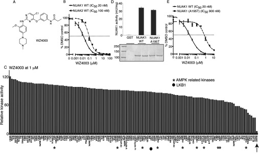
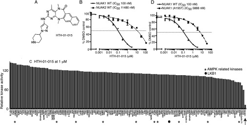
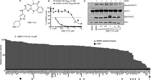
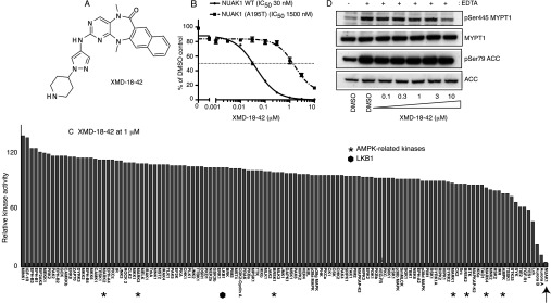
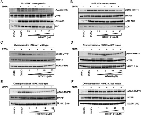
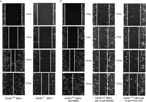
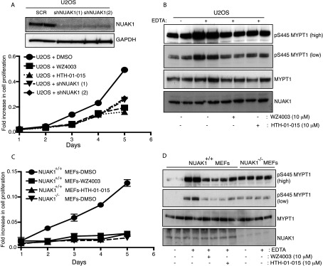
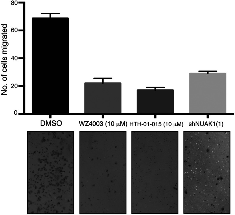
The related NUAK1 and NUAK2 are members of the AMPK (AMP-activated protein kinase) family of protein kinases that are activated by the LKB1 (liver kinase B1) tumour suppressor kinase. Recent work suggests they play important roles in regulating key biological processes including Myc-driven tumorigenesis, senescence, cell adhesion and neuronal polarity. In the present paper we describe the first highly specific protein kinase inhibitors of NUAK kinases namely WZ4003 and HTH-01-015. WZ4003 inhibits both NUAK isoforms (IC50 for NUAK1 is 20 nM and for NUAK2 is 100 nM), whereas HTH-01-015 inhibits only NUAK1 (IC50 is 100 nM). These compounds display extreme selectivity and do not significantly inhibit the activity of 139 other kinases that were tested including ten AMPK family members. In all cell lines tested, WZ4003 and HTH-01-015 inhibit the phosphorylation of the only well-characterized substrate, MYPT1 (myosin phosphate-targeting subunit 1) that is phosphorylated by NUAK1 at Ser(445). We also identify a mutation (A195T) that does not affect basal NUAK1 activity, but renders it ~50-fold resistant to both WZ4003 and HTH-01-015. Consistent with NUAK1 mediating the phosphorylation of MYPT1 we find that in cells overexpressing drug-resistant NUAK1[A195T], but not wild-type NUAK1, phosphorylation of MYPT1 at Ser(445) is no longer suppressed by WZ4003 or HTH-01-015. We also demonstrate that administration of WZ4003 and HTH-01-015 to MEFs (mouse embryonic fibroblasts) significantly inhibits migration in a wound-healing assay to a similar extent as NUAK1-knockout. WZ4003 and HTH-01-015 also inhibit proliferation of MEFs to the same extent as NUAK1 knockout and U2OS cells to the same extent as NUAK1 shRNA knockdown. We find that WZ4003 and HTH-01-015 impaired the invasive potential of U2OS cells in a 3D cell invasion assay to the same extent as NUAK1 knockdown. The results of the present study indicate that WZ4003 and HTH-01-015 will serve as useful chemical probes to delineate the biological roles of the NUAK kinases.


Figures

Figure 1
Figure 1. WZ4003, a specific NUAK1 and NUAK2 inhibitor
(A) Chemical structure of the NUAK1/NUAK2 inhibitor WZ4003. (B) Wild-type (WT) GST–NUAK1 and GST–NUAK2 were assayed using 200 μM Sakamototide in the presence of 100 μM [γ-32P]ATP (~500 c.p.m./pmol) with the indicated concentrations of WZ4003. The IC50 graph was plotted using GraphPad Prism software with non-linear regression analysis. The results are presented as the percentage of kinase activity relative to the DMSO-treated control. Results are means±S.D. for triplicate reactions with similar results obtained in at least one other experiment. (C) Kinase profiling of the WZ4003 inhibitor at 1 μM was carried out against the panel of 140 kinases at the The International Centre for Protein Kinase Profiling (http://www.kinase-screen.mrc.ac.uk/). AMPK family kinases are indicated with an asterisk, LKB1 with a filled hexagon and NUAK1 with an arrow. The full names of the kinases can be found in the legend to Supplementary Table S1 (at http://www.biochemj.org/bj/457/bj4570215add.htm). (D) Wild-type (WT) GST–NUAK1 and GST–NUAK1[A195T] were purified from HEK-293 cells following transient transfection and relative levels of wild-type and mutant enzymes were analysed by Coomassie Blue staining of a polyacrylamide gel (bottom panel). Intrinsic kinase activities of the equivalent amounts of NUAK1 and NUAK1[A195T] were compared by carrying out a quantitative kinase activity assay by calculating the relative kinase-mediated incorporation of [γ-32P]ATP into the Sakamototide substrate peptide. Values are means±S.D. for an experiment carried out in triplicate. (E) As in (B) except that WZ4003 comparative IC50 values were derived for wild-type (WT) GST–NUAK1 and GST—NUAK1[A195T].
Figure 2
Figure 2. HTH-01-015, a specific NUAK1 inhibitor
(A) Chemical structure of the NUAK1-specific inhibitor HTH-01-015. (B) Wild-type (WT) GST–NUAK1 and GST–NUAK2 were assayed using 200 μM Sakamototide in the presence of 100 μM [γ-32P]ATP (~500 c.p.m./pmol) with the indicated concentrations of HTH-01-015. The IC50 graph was plotted using Graphpad Prism software with non-linear regression analysis. The results are presented as the percentage of kinase activity relative to the DMSO-treated control. Results are means±S.D. for triplicate reactions with similar results obtained in at least one other experiment. (C) Kinase profiling of the HTH-01-015 inhibitor at 1 μM was carried out against the panel of 140 kinases at the The International Centre for Protein Kinase Profiling (http://www.kinase-screen.mrc.ac.uk/). AMPK family kinases are indicated with an asterisk, LKB1 with a filled hexagon and NUAK1 with an arrow. The full names of the kinases can be found in the legend to Supplementary Table S1 (at http://www.biochemj.org/bj/457/bj4570215add.htm). (D) As in (B) except that HTH-01-015 comparative IC50 values were derived for wild-type (WT) GST–NUAK1 and GST–NUAK1[A195T].
Figure 3
Figure 3. XMD-17-51, a potent semi-specific NUAK1 inhibitor
(A) Chemical structure of XMD-17-51. (B) Wild-type (WT) GST–NUAK1 and GST–NUAK1[A195T] were assayed using 200 μM Sakamototide in the presence of 100 μM [γ-32P]ATP (~500 c.p.m./pmol) with the indicated concentrations of XMD-17-51. The IC50 graph was plotted using Graphpad Prism software with non-linear regression analysis. The results are presented as the percentage of kinase activity relative to the DMSO-treated control. Results are means±S.D. for triplicate reactions with similar results obtained in at least one other experiment. (C) Kinase profiling of the XMD-17-51 inhibitor at 1 μM was carried out against the panel of 140 kinases at the The International Centre for Protein Kinase Profiling (http://www.kinase-screen.mrc.ac.uk/). AMPK family kinases are indicated with an asterisk, LKB1 with a filled hexagon and NUAK1 with an arrow. The full names of the kinases can be found in the legend to Supplementary Table S1 (at http://www.biochemj.org/bj/457/bj4570215add.htm). (D) HEK-293 cells were treated in the absence (DMSO) or presence of the indicated concentrations of XMD-17-51 over 16 h. Cell medium was then replaced with either normal DMEM containing no EDTA-PBS-based cell dissociation buffer (−) or EDTA-PBS-based cell dissociation buffer (+) containing the same concentration of XMD-17-51 that the cells were previously incubated in. Cell detachment was induced with gentle tapping of the plates followed by gentle centrifugation at 70 g for 3 min. Cells were lysed immediately after removal of the supernatant. Endogenous MYPT1 was immunoprecipitated from 0.5 mg of the cell lysates. The immunoprecipitates were immunoblotted for the detection of p-Ser445 MYPT1 and total MYPT1. The cell lysates were subjected to immunoblotting for the detection of p-Ser79 ACC and total ACC. Similar results were obtained in three separate experiments.
Figure 4
Figure 4. XMD-18-42, a semi-specific NUAK1 inhibitor
(A) Chemical structure of XMD-18-42. (B) Wild-type (WT) GST–NUAK1 and GST–NUAK1[A195T] were assayed using 200 μM Sakamototide in the presence of 100 μM [γ-32P]ATP (~500 c.p.m./pmol) with the indicated concentrations of XMD-18-42. The IC50 graph was plotted using Graphpad Prism software with non-linear regression analysis. The results are presented as the percentage of kinase activity relative to the DMSO-treated control. Results are means±S.D. for triplicate reactions with similar results obtained in at least one other experiment. (C) Kinase profiling of the XMD-18-42 inhibitor at 1 μM was carried out against the panel of 140 kinases at the The International Centre for Protein Kinase Profiling (http://www.kinase-screen.mrc.ac.uk/). AMPK family kinases are indicated with an asterisk, LKB1 with a filled hexagon and NUAK1 with an arrow. The full names of the kinases can be found in the legend to Supplementary Table S1 (at http://www.biochemj.org/bj/457/bj4570215add.htm). (D) HEK-293 cells were treated in the absence (DMSO) or presence of the indicated concentrations of XMD-18-42 over 16 h. Cell medium was then replaced with either normal DMEM containing no EDTA-PBS-based cell dissociation buffer (−) or EDTA-PBS-based cell dissociation buffer (+) containing the same concentration of XMD-18-42 that the cells were previously incubated in. Cell detachment was induced with gentle tapping of the plates followed by gentle centrifugation at 70 g for 3 min. Cells were lysed immediately after removal of the supernatant. Endogenous MYPT1 was immunoprecipitated from 0.5 mg of the cell lysates. The immunoprecipitates were immunoblotted for the detection of p-Ser445 MYPT1 and total MYPT1. The cell lysates were subjected to immunoblotting for the detection of p-Ser79 ACC and total ACC. Similar results were obtained in three separate experiments.
Figure 5
Figure 5. HTH-01-015 and WZ4003 inhibit MYPT1 Ser445 phosphorylation in vivo
(A) HEK-293 cells were treated in the absence (DMSO) or presence of the indicated concentrations of WZ4003 over 16 h. Cell medium was then replaced with either normal DMEM containing no EDTA-PBS-based cell dissociation buffer (−) or EDTA-PBS-based cell dissociation buffer (+) containing the same concentration of WZ4003 that the cells were previously incubated in. Cell detachment was induced with gentle tapping of the plates followed by gentle centrifugation at 70 g for 3 min. Cells were lysed immediately after removal of the supernatant. Endogenous MYPT1 was immunoprecipitated from 0.5 mg of the cell lysates. The immunoprecipitates were immunoblotted for the detection of p-Ser445 MYPT1 and total MYPT1. The cell lysates were subjected to immunoblotting for the detection of p-Ser79 ACC and total ACC. Similar results were obtained in three separate experiments. (B) As in (A) except for the HTH-01-015 inhibitor was used. (CF) As above except that HEK-293 Flp/In T-Rex cells stably expressing the indicated wild-type HA-tagged NUAK1 or drug-resistant HA-tagged NUAK1[A195T] were used. Similar results were obtained in three separate experiments for all data shown on this Figure.
Figure 6
Figure 6. NUAK1 inhibition suppresses cell migration
(A) NUAK1+/+ and NUAK1−/− MEFs were split into the chambers (as described in the Materials and methods section). The inserts were then removed and a wound-healing assay was carried out in triplicate. Snapshots at specific time points from time-lapse microscopy were used as representative images for comparison between the migration properties of NUAK1+/+ and NUAK1−/− MEFs. (B) The migration assay of NUAK1+/+ MEFs treated with or without 10 μM WZ4003 or HTH-01-015 was carried out as in (A).
Figure 7
Figure 7. NUAK1 inhibition suppresses cell proliferation
(A) U2OS cells were incubated with or without 10 μM WZ4003 or 10 μM HTH-01-015 and a cell proliferation assay was carried out over 5 days in triplicate using the CellTiter 96® AQueous Non-Radioactive Cell Proliferation Assay kit (Promega) (as described in the Materials and methods section). U2OS cells in which NUAK1 has been knocked-down using two different shRNA hairpins were used in parallel as controls. The efficiency of the knock down of each shRNA is shown in top panel. SCR, control scrambled shRNA hairpin; shNUAK1 (1), first NUAK1 shRNA hairpin; shNUAK1 (2), second NUAK1 shRNA hairpin. (B) U2OS cells were treated with (+) or without (−) 10 μM WZ4003 or 10 μM HTH-01-015. After 16 h cell media was removed and cells were treated with EDTA-PBS-based cell dissociation buffer supplemented with 10 μM WZ4003, 10 μM HTH-01–015 or DMSO for 20 min. Cell detachment was induced with gentle tapping of the plates followed by gentle centrifugation at 70 g for 3 min. Cells were lysed immediately after removal of the media and immunoblotted for the detection of the indicated antibodies. (C and D) As above, except NUAK1+/+ and NUAK1−/− MEFs were used. Similar results were obtained in three separate experiments.
Figure 8
Figure 8. NUAK1 inhibition suppresses invasion potential
U2OS cells treated without (DMSO) or with 10 μM WZ4003 or 10 μM HTH-01-015 were plated on to a Transwell® invasion assay plate in triplicate using 10% FBS as a chemoattractant. Cells that had invaded through to the lower face of the filters were fixed and photographed (×10 magnification). The cells that migrated were counted and the data are presented as the mean number of migrated cells±S.D. U2OS cells with NUAK1-knockdown were used in parallel as a control. Similar results were obtained in two separate experiments with each condition analysed in triplicate.

References

    1. Lizcano J. M., Goransson O., Toth R., Deak M., Morrice N. A., Boudeau J., Hawley S. A., Udd L., Makela T. P., Hardie D. G., Alessi D. R. LKB1 is a master kinase that activates 13 kinases of the AMPK subfamily, including MARK/PAR-1. EMBO J. 2004;23:833–843. - PMC - PubMed
    1. Alessi D. R., Sakamoto K., Bayascas J. R. LKB1-dependent signaling pathways. Annu. Rev. Biochem. 2006;75:137–163. - PubMed
    1. Jaleel M., McBride A., Lizcano J. M., Deak M., Toth R., Morrice N. A., Alessi D. R. Identification of the sucrose non-fermenting related kinase SNRK, as a novel LKB1 substrate. FEBS Lett. 2005;579:1417–1423. - PubMed
    1. Hardie D. G., Ross F. A., Hawley S. A. AMPK: a nutrient and energy sensor that maintains energy homeostasis. Nat. Rev. Mol. Cell Biol. 2012;13:251–262. - PMC - PubMed
    1. Matenia D., Mandelkow E. M. The tau of MARK: a polarized view of the cytoskeleton. Trends Biochem. Sci. 2009;34:332–342. - PubMed

Publication types

MeSH terms