. 2014 Mar 1;23(5):502-14.
doi: 10.1089/scd.2013.0274. Epub 2013 Dec 4.

Adult human nasal mesenchymal-like stem cells restore cochlear spiral ganglion neurons after experimental lesion

Affiliations

Adult human nasal mesenchymal-like stem cells restore cochlear spiral ganglion neurons after experimental lesion

Esperanza Bas et al. Stem Cells Dev. .

Abstract

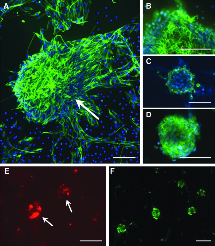
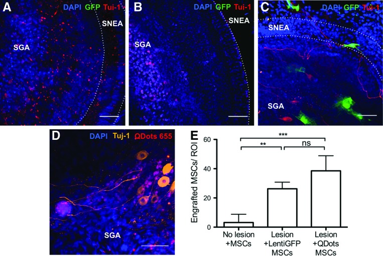
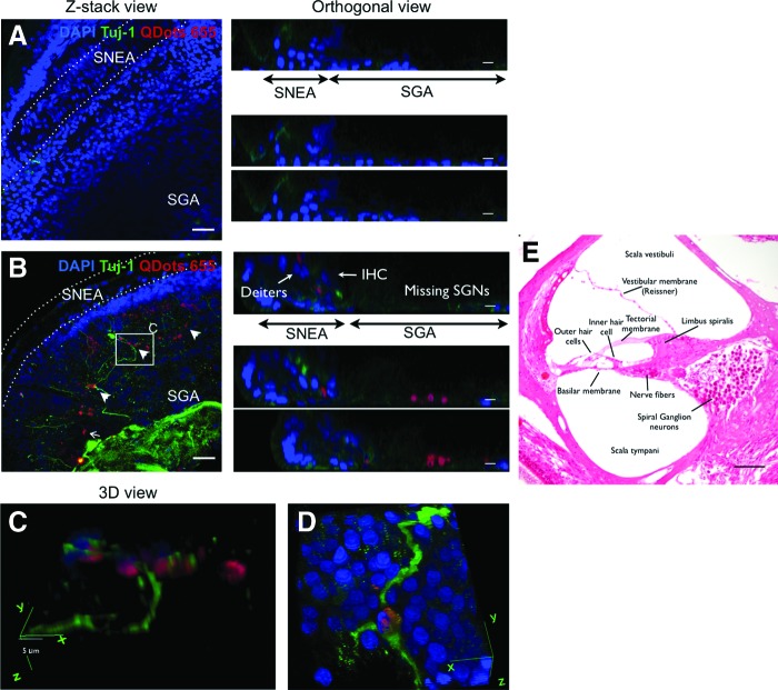
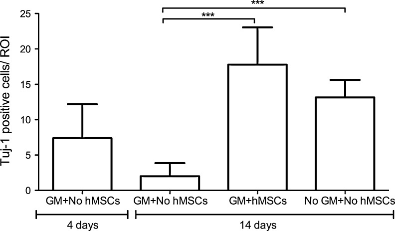
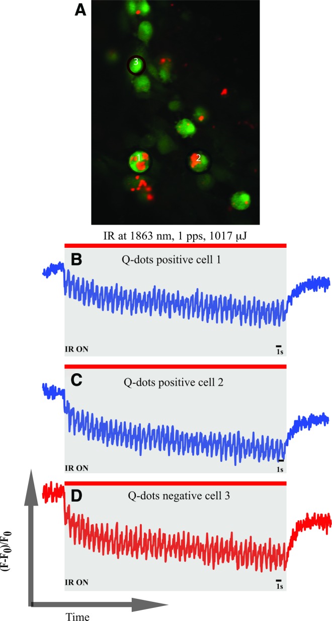
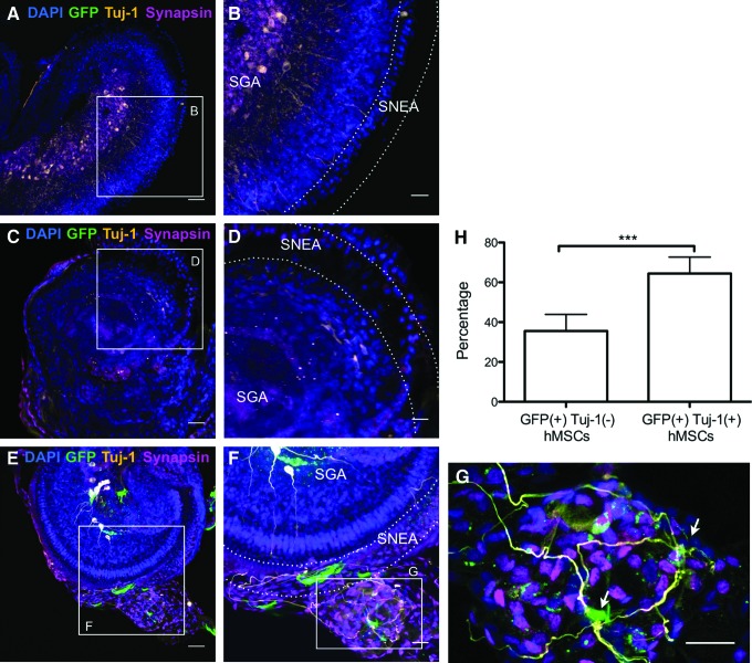
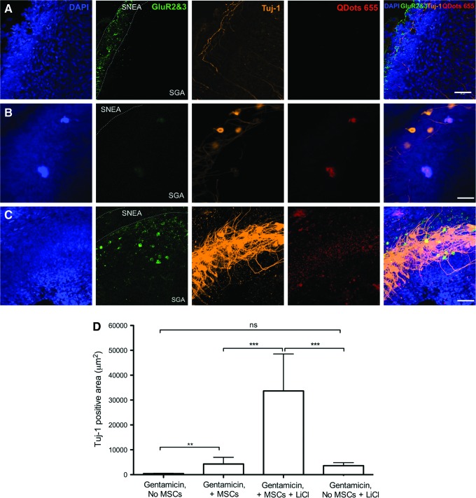
A loss of sensory hair cells or spiral ganglion neurons from the inner ear causes deafness, affecting millions of people. Currently, there is no effective therapy to repair the inner ear sensory structures in humans. Cochlear implantation can restore input, but only if auditory neurons remain intact. Efforts to develop stem cell-based treatments for deafness have demonstrated progress, most notably utilizing embryonic-derived cells. In an effort to bypass limitations of embryonic or induced pluripotent stem cells that may impede the translation to clinical applications, we sought to utilize an alternative cell source. Here, we show that adult human mesenchymal-like stem cells (MSCs) obtained from nasal tissue can repair spiral ganglion loss in experimentally lesioned cochlear cultures from neonatal rats. Stem cells engraft into gentamicin-lesioned organotypic cultures and orchestrate the restoration of the spiral ganglion neuronal population, involving both direct neuronal differentiation and secondary effects on endogenous cells. As a physiologic assay, nasal MSC-derived cells engrafted into lesioned spiral ganglia demonstrate responses to infrared laser stimulus that are consistent with those typical of excitable cells. The addition of a pharmacologic activator of the canonical Wnt/β-catenin pathway concurrent with stem cell treatment promoted robust neuronal differentiation. The availability of an effective adult autologous cell source for inner ear tissue repair should contribute to efforts to translate cell-based strategies to the clinic.


Figures

<b>FIG. 1.</b>
FIG. 1.
Human nasal mesenchymal-like stem cells (nasal MSCs) express an appropriate MSC phenotype. (A) Under sphere-forming conditions, cells express the intermediate filament Nestin (green); arrow indicates a large nestin (+) sphere. The surface markers characteristic of MSCs are also expressed, including CD90 (B), CD105 (C), and STRO1 (D). Nuclei are counterstained with 4′,6-diamidino-2-phenylindole (DAPI) (blue). (E, F) Live nasal MSCs were labeled with either fluorescent nanospheres (E, Qtracker 655; Molecular Probes Eugene, OR, USA) or green fluorescent protein (GFP) via lentiviral tranfection (F) to facilitate tracking cells in a co-culture with rat cochlea. Bar=50 μm. Color images available online at www.liebertpub.com/scd
<b>FIG. 2.</b>
FIG. 2.
Human nasal MSCs engraft in lesioned rat cochleae. Co-cultures with lentiGFP-transfected MSCs are shown in (A–C) (green); cultures with QDot 655-labeled MSCs are shown in (D) (red). The specimens were co-stained with Tuj-1 [red in (AC) and orange in (D)] as a neuron-specific class III beta-tubulin marker and DAPI to visualize nuclei (blue). (A) Un-lesioned cultures without stem cell treatment show remnants of neurites after a total of 14 days in culture. (B) Gentamicin (GM)-lesioned cochlear cultures in which nasal MSCs were not added show absence of neurons and shrunken nuclei. (C, D) GM-lesioned explants co-cultured with nasal MSCs show healthy neurons and engrafted nasal MSCs. The sensorineural epithelia area (SNEA) and spiral ganglion area (SGA) are indicated in each photograph. Very few nasal MSCs were identifiable into control, un-lesioned cultures that were stem cell treated [(E), microphotograph not included], indicating that injury or lesion permits stem cell engraftment: 38.5±4.2 QDot-labeled MSCs and 26.2±2.3 LentiGFP-transfected MSCs were identifiable in lesioned cultures; 3.2±2.3 MSCs were identifiable in un-lesioned cultures (n=6 independent specimens/group). Plotted data correspond to the mean±standard deviation (SD). Analysis of variance (ANOVA) followed by Tukey's test was used for statistics. **P<0.05, ***P<0.005, ns, not significant. Bar=50 μm. Color images available online at www.liebertpub.com/scd
<b>FIG. 3.</b>
FIG. 3.
Engrafted nasal MSCs migrate to the spiral ganglion region and promote an increase in spiral ganglion neurons. GM-lesioned cochleae in culture were either untreated (A) or treated with nasal MSCs [(BD), red]. An analysis after 14 days shows that cultures which did not receive nasal MSCs have largely absent Tuj-1 neuronal staining in the spiral ganglion (A); in MSC-treated cultures, the nasal MSCs tend to cluster in the lesioned spiral ganglion [(B), arrows]; this is readily apparent on orthogonal views in (B), where the Deiter's cells and inner hair cells are identified in the sensorineural epithelia area, and the red labeled MSCs engraft in the spiral ganglion area. There are prominent Tuj-1-labeled neuronal processes extending from this region in MSC-treated cultures. Nuclei are counterstained with DAPI (blue); bar=50 μm in Z-stack views and 25 μm in orthogonal views. (C, D) A three-dimensional view from a boxed region in (B) (C) and a similar region from a different specimen (D) show how some neurons with Tuj-1 (+) processes (green) are labeled with Qdot beads (red), indicating that they are human nasal MSC derived; bar=5 μm; 40×oil immersion objective. (E) Eosin-stained histologic section through the cochlea, labeled to provide orientation for confocal figures. SGNs, spiral ganglion neurons; IHC, inner hair cell. Arrowheads indicate TuJ-1 (+) nasal MSC-derived cells. Color images available online at www.liebertpub.com/scd
<b>FIG. 4.</b>
FIG. 4.
Human nasal mesenchymal stem cell (hMSC) treatment restores spiral ganglion cell population. Quantification of TuJ-1-expressing neurons in the spiral ganglion from GM-lesioned cultures with no stem cell treatment (GM+No hMSCs), GM-lesioned cultures with human nasal MSC treatment (GM+hMSCs), or un-lesioned cultures (No GM+No hMSCs) indicates that there is an ongoing loss of neurons from 4 to 14 days after GM lesion in cultures not treated with stem cells, and that hMSC treatment results in a restoration of the neuronal population. Mean neuronal counts are markedly increased in GM-lesioned cultures that received hMSCs (17.8±5.1; n=14 cultures) versus GM-lesioned cultures which did not receive hMSCs (2.0±1.8; n=8 cultures, ***P<0.005). Plotted data correspond to the mean±SD. ANOVA followed by Tukey's test was used for statistical analysis.
<b>FIG. 5.</b>
FIG. 5.
MSC-derived cells display excitable responses to infrared laser stimulus. (A) Shows QDot-positive and -negative cells after loading with Ca2+-sensitive dye Fluo-4 AM. Two QDot-positive (ie, nasal MSC-derived) cells and one QDot-negative cell were selected to analyze their responses to infrared radiation (IR) stimulation for 1 min at 1,893 nm, 1 pps, and 1,017 mJ of radiant energy. Normalized fluorescence values were plotted over time. Traces (B) and (C) show the IR-evoked [Ca2+]i transients in the two selected QDot-positive cells. Trace (D) shows the response of a QDot-negative endogenous cell, which can be a spiral ganglion neuron or a glial cell. Responses obtained in traces (BD) are similar to the ones obtained in excitable cells such as neurons or cardiomyocytes. Color images available online at www.liebertpub.com/scd
<b>FIG. 6.</b>
FIG. 6.
Differentiation of spiral ganglion neurons in human nasal MSC-treated cochlear cultures. (A, B) Un-lesioned control at 1 day post-explantation shows extensive synapsin (magenta) and Tuj-1 (orange) labeling. (C, D) Neuron-specific Tuj-1 labeling and synapsin staining is absent in GM-lesioned cochlea without stem cell treatment. (EG) GM-lesioned stem cell-treated cultures exhibit robust Tuj-1 labeling (orange) of neurons and synapsin labeling (magenta) among lentiGFP-transfected nasal MSCs (green); SNEA and SGA are indicated in the figures. A cluster of ectopically-located nasal MSC-derived cells is visible in this field (G), with extensive synapsin expression and several prominent TuJ-1 (+) processes extending toward the sensory epithelium. Arrows indicate GFP (+)/TuJ-1 (+) cells. (A, C, E) Bar=50 μm. (B, D, F, G) Bar=25 μm. (H) Graph showing the percentage of GFP-labeled MSCs that are positive and negative for Tuj-1 staining. Plotted data correspond to mean±SD. Unpaired two-tailed t-test was applied (n=6 independent specimens/group, ***P<0.001). Color images available online at www.liebertpub.com/scd
<b>FIG. 7.</b>
FIG. 7.
Pharmacologic activation of Wnt signaling pathway alters the neurite morphology in stem cell-treated cultures at 14 days. (A) Control un-lesioned cultures show typical Tuj-1 labeling, along with low levels of glutamate receptor GluR2/3 when maintained in vitro for 14 days. (B) In GM-lesioned cultures treated with human nasal MSCs alone, both TuJ-1 (+) neurons (orange) and faint GluR2/3 staining (green) are evident. (C) In GM-lesioned cultures treated with stem cells and LiCl (a Wnt/β-catenin pathway activator), TuJ-1 (+) cells exhibit pronounced density of staining and dendritic arborization (orange label, middle column); GluR2/3 staining is also more robust (green) in adjacent areas; SNEA and SGA are labeled; bar=50 μm. (D) Measurement of the TuJ-1 labeled area, which labels neurites, indicates a significant increase in stem cell-treated cultures that are maintained with LiCl. The TuJ-1 label reflects the increased complexity and density of neurite outgrowth. **P<0.05, ***P<0.005, ns, not significant. Color images available online at www.liebertpub.com/scd

References

    1. Pittenger MF. (1999). Multilineage potential of adult human mesenchymal stem cells. Science 284:143–147 - PubMed
    1. Williams AR. and Hare JM. (2011). Mesenchymal stem cells: biology, pathophysiology, translational findings, and therapeutic implications for cardiac disease. Circ Res 109:923–940 - PMC - PubMed
    1. Hatzistergos KE,, Quevedo H, Oskouei BN, Hu Q, Feigenbaum GS, Margitich IS, Mazhari R, Boyle AJ, Zambrano JP, et al. (2010). Bone marrow mesenchymal stem cells stimulate cardiac stem cell proliferation and differentiation. Circ Res 107:913–922 - PMC - PubMed
    1. Murrell W, Feron F, Wetzig A, Cameron N, Splatt K, Bellette B, Bianco J, Perry C, Lee G. and Mackay-Sim A. (2005). Multipotent stem cells from adult olfactory mucosa. Dev Dyn 233:496–515 - PubMed
    1. Jakob M, Hemeda H, Janeschik S, Bootz F, Rotter N, Lang S. and Brandau S. (2010). Human nasal mucosa contains tissue-resident immunologically responsive mesenchymal stromal cells. Stem Cells Dev 19:635–644 - PubMed

Publication types