. 2014 Jul;171(14):3339-51.
doi: 10.1111/bph.12493.

Short-term exposure to oleandrin enhances responses to IL-8 by increasing cell surface IL-8 receptors

Affiliations

Short-term exposure to oleandrin enhances responses to IL-8 by increasing cell surface IL-8 receptors

Nune Raviprakash et al. Br J Pharmacol. 2014 Jul.

Abstract

Background and purpose: One of the first steps in host defence is the migration of leukocytes. IL-8 and its receptors are a chemokine system essential to such migration. Up-regulation of these receptors would be a viable strategy to treat dysfunctional host defence. Here, we studied the effects of the plant glycoside oleandrin on responses to IL-8 in a human monocytic cell line.

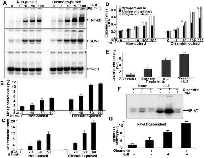
Experimental approach: U937 cells were incubated with oleandrin (1-200 ng mL(-1) ) for either 1 h (pulse) or for 24 h (non-pulse). Apoptosis; activation of NF-κB, AP-1 and NFAT; calcineurin activity and IL-8 receptors (CXCR1 and CXCR2) were measured using Western blotting, RT-PCR and reporter gene assays.

Key results: Pulse exposure to oleandrin did not induce apoptosis or cytoxicity as observed after non-pulse exposure. Pulse exposure enhanced activation of NF-κB induced by IL-8 but not that induced by TNF-α, IL-1, EGF or LPS. Exposure to other apoptosis-inducing compounds (azadirachtin, resveratrol, thiadiazolidine, or benzofuran) did not enhance activation of NF-κB. Pulse exposure to oleandrin increased expression of IL-8 receptors and chemotaxis, release of enzymes and activation of NF-κB, NFAT and AP-1 along with increased IL-8-mediated calcineurin activation, and wound healing. Pulse exposure increased numbers of cell surface IL-8 receptors.

Conclusions and implications: Short-term (1 h; pulse) exposure to a toxic glycoside oleandrin, enhanced biological responses to IL-8 in monocytic cells, without cytoxicity. Pulse exposure to oleandrin could provide a viable therapy for those conditions where leukocyte migration is defective.

Keywords: IL-8 receptor; NF-κB; apoptosis; cardiac glycoside; chemotaxis.


Figures

Figure 1
Figure 1
Effect of pulsed and non-pulsed exposure to oleandrin on cell viability. U-937 cells were treated with different concentrations of oleandrin for 1 h and then cells were washed and cultured for a further 24 h; this is referred to as ‘pulse exposure’. In another set, U-937 cells were incubated with the same concentrations of oleandrin for 24 h (non-pulse exposure). In A, the MTT assay was used to measure cell viability; data from one of three experiments is shown. In B, cell death was assessed by nuclear fragmentation assay as detected by PI staining, following the same pulse and non-pulse exposures,. In (C), another measure of apoptosis, cleaved PARP, in whole cell extracts of U-937 cells after pulse and non-pulse exposures to oleandrin, was detected by Western blot. (D) Activities of caspase 3 and 8 were measured in whole cell extracts. Results are expressed as fold activation, relative to values in cells without oleandrin exposure. U-937 cells were incubated with oleandrin (100 ng mL−1) under pulsed or non-pulsed conditions, followed by washing and culturing for different times. They were then stimulated with IL-8 (100 ng mL−1) for 2 h. Nuclear extracts were prepared and assayed for NF-κB DNA binding (E). Cell death was detected by PI staining of cell nuclei (F) and viability (G) by the MTT assay.
Figure 2
Figure 2
Effect of pulse exposure to a range of apoptosis–inducing compounds on IL-8- or TNF-induced NF-κB activation. In (A), U-937 cells were pulsed with azadirachtin (10 μM), benzofuran (100 nM), oleandrin (100 ng mL−1), P3-25 (100 nM), or resveratrol (10 μM) for 1h then washed and cultured in drug-free media for another 24h. Cells were then were stimulated with TNF (100 pM) or IL-8 (100 ng mL−1) for 2 h. As an indication of cell activation, binding of NF-κB to DNA was measured in nuclear extracts. In (B), U-937 cells were transfected with NF-κB-luciferase and GFP constructs; 45% of the cells were GFP positive. Cells were treated with the different compounds for 1 h, washed and cultured for 24 h. Whole cell extracts were prepared and luciferase activity was measured and expressed as fold of values in untreated cells (set to unity).
Figure 3
Figure 3
Effect of oleandrin pulse on NF-κB activation mediated by different agonists and in different cells. In (A), oleandrin-pulsed U-937 cells were stimulated with TNF (10 pM), LPS (100 ng mL−1), IL-1 (50 nM), IL-8 (50 ng mL−1) or EGF (100 pM) for 6 h. NF-κB DNA binding was assayed in nuclear extracts (A). In (B), a range of different cell types was pulsed with oleandrin and then stimulated with 10 and 50 ng mL−1 IL-8 for 4 h and NF-κB DNA binding was assayed.
Figure 4
Figure 4
Effect of oleandrin pulse on IL-8-mediated signalling pathway. In (A), Oleandrin-pulsed U-937 cells, incubated with antibodies to the two IL-8 receptors (1 μg mL−1 each) for 2 h, were stimulated with IL-8 for 4 h. NF-κB DNA binding was assayed in nuclear extracts. In (B), Oleandrin-pulsed cells were cultured for 12 h, transfected with 1 μg of TRAF6-DN or (in C) the TRAF6 construct for 3 h, washed and cultured for 12 h. Cells were then stimulated with 100 ng mL−1 IL-8 for 4 h. NF-κB DNA binding was measured. In (D), oleandrin-pulsed U-937 cells were incubated with 200 μM of TRAF6-BP or TRAF6-BP (Mut) for 4 h and then stimulated with IL-8 for 4 h. NF-κB DNA binding was determined in nuclear extracts. In (E), oleandrin-pulsed cells were stimulated with NGF (100 nM), FMLP (100 nM), α-MSH (1 μM), vasopressin (100 nM), serotonin (100 nM) or IL-8 (100 ng mL−1) for 6 h. NF-κB DNA binding was measured in nuclear extracts.
Figure 5
Figure 5
Effect of oleandrin pulse on expression of IL-8 receptors. In (A), total RNA was isolated from cells pulsed with oleandrin (1h) followed by culture in oleandrin-free medium for 24 h. The amounts of mRNA for CXCR4, CXCR2, CXCR1 and actin were measured by RT-PCR. In (B), the amount of CXCR1 on U 937 cells was detected by immunofluorescence using an appropriate antibody and visualised with goat anti-rabbit IgG conjugated with Alexa fluor 594. (C) Cells were pulsed with oleandrin for 1 h, then washed and treated with 1 μM cycloheximide (CHX) or 10 μM cystamine for 1 h and then cultured for 24 h. The amount of IL-8 receptors were determined in 100 μg of whole cell extracts (WCE) and 50 μg of membrane extract by Western blot. In (D), U-937 cells, either non-pulsed or pulsed with oleandrin, treated with CHX, cystamine or oleandrin for 2 h were cultured for 24 h. Cells were stimulated with 100 ng mL−1 IL-8 for 4 h. NF-κB activation was assayed in nuclear extracts.
Figure 6
Figure 6
Effect of oleandrin pulse on IL-8-mediated biological responses in U-937 cells. (A) Oleandrin-pulsed and non-pulsed cells were stimulated with different concentrations of IL-8 for 4 h. Activation of NF-κB, AP-1 and Oct1 was determined in nuclear extracts. (B) Oxidative burst in cells stimulated by IL-8 was assessed with nitroblue tetrazolium (NBT) dye. The proportion of NBT positive cells was determined and shown as fold of of values in untreated cells (set to unity). (C) Migration of U-937 cells to IL-8 was determined in Boyden chambers and the chemotactic index (ratio of cell migration with and without IL-8) is shown. (D) Activities of myeloperoxidase, β-D-glucuronidase and alkaline phosphatase were measured in the culture supernatant of oleandrin-pulsed and non-pulsed cells, stimulated with different concentrations of IL-8 for 4 h. Activities are expressed as the OD values corresponding to each assay. In (E), the calcineurin activity in oleandrin-pulsed cells stimulated with IL-8 (100 ng mL−1) for 2 h is shown. (F) Oleandrin-pulsed cells, incubated with cyclosporine A (CsA; 2.5 μM) for 2 h were stimulated with IL-8 and NFAT activation was measured in nuclear extracts. In (G), cells were pulsed with oleandrin for 1 h, cultured for 12 h, and then transfected with NFAT-luciferase construct (1 μg) for 3 h and cultured for another 12 h. Cells were then stimulated with IL-8 (100 ng mL−1) for 12 h and the luciferase activity was measured.
Figure 7
Figure 7
Effect of oleandrin exposure on a model of wound healing. Cultures of A549 cells at 50% confluency were pulsed or non-pulsed with oleandrin. The cell monolayer was then scratched with a sterile needle to simulate the wound and the cultures stimulated with 50 or 100 ng mL−1 of IL-8. Phase contrast images were taken at different times after wounding, as indicated in the Figure.
Figure 8
Figure 8
Effect of lipid compounds on the effects of pulse exposure to oleandrin. (A) U-937 cells were incubated with cholesterol, cephalin, sphingosine or lecithin (500 ng mL−1 each) for 4 h and then pulsed with oleandrin (1h), washed and incubated for a further 24h. Non-pulsed cells were similarly exposed to the lipids and then incubated with oleandrin for 24h. Cells were then stimulated with IL-8 for 4 h and NF-κB activation was assessed in nuclear extracts. (B) Cells, incubated with lipids for 4 h were pulsed or non-pulsed with oleandrin. For the last 2 h, cells were treated with oleandrin (100 ng mL−1) in one set of samples, followed by stimulation with IL-8 for 4 h and NF-κB activation assayed. (C) Cells were pulsed with a range of concentrations of oleandrin (10–200 ng mL−1). They were then stimulated with 50 ng mL−1 phorbol myristate acetate (PMA) for 2 h. Activation of NFAT was determined in nuclear extracts and IL-8 receptors were measured in whole cell extracts (WCE). (D) Cells, incubated for 4 h with the combination of lipids were pulsed or non-pulsed with oleandrin. NFAT activation was measured. IL-8 receptors were measured by Western blot in whole cell extracts. (E) Cells pulsed with oleandrin for 1 h and then cultured with pyrrolidine dithiocarbamate (PDTC) (100 μM), BAPTA-AM (2.5 μM), or cyclosporine A (CsA) (2.5 μM) for 24 h. In one set, cells were treated with 100 ng mL−1 oleandrin for 4 h. NFAT activation was assayed in nuclear extracts.

Similar articles

Cited by

References

    1. Abbasi S, Su B, Kellems RE, Yang J, Xia Y. The essential role of MEKK3 signaling in angiotensin II-induced calcineurin/nuclear factor of activated T-cells activation. J Biol Chem. 2005;280:36737–36746. - PubMed
    1. Alexander SPH, Benson HE, Faccenda E, Pawson AJ, Sharman JL, Spedding M, et al. The Concise Guide to PHARMACOLOGY 2013/14: G Protein-Coupled Receptors. Br J Pharmacol. 2013;170:1459–1581. - PMC - PubMed
    1. Bidyasar S, Kurzrock R, Falchook GS, Naing A, Wheler JJ, Durand J, et al. A first-in-human phase I trial of PBI-05204 (oleandrin), an inhibitor of Akt, FGF-2, NF-kB, and p70S6K in advanced solid tumor patients. J Clin Oncol. 2009;27:15S. Abst 3537.
    1. Crabtree GR, Olson EN. NFAT signaling: choreographing the social lives of cells. Cell. 2002;109(Suppl):S67–S79. - PubMed
    1. Kingsbury TJ, Cunningham KW. A conserved family of calcineurin regulators. Genes Dev. 2000;14:1595–1604. - PMC - PubMed

Publication types

LinkOut - more resources