

T1ρ magnetic resonance imaging for detection of early cartilage changes in knees of asymptomatic collegiate female impact and nonimpact athletes
- PMID: 24172654
- PMCID: PMC6425943
- DOI: 10.1097/JSM.0000000000000013
T1ρ magnetic resonance imaging for detection of early cartilage changes in knees of asymptomatic collegiate female impact and nonimpact athletes
Abstract
Objective: To determine if T1ρ magnetic resonance imaging (T1ρ MRI) could assess early articular cartilage changes in knees of asymptomatic female collegiate athletes. It was hypothesized that impact cohort would demonstrate greater changes than nonimpact cohort.
Design: An institutional review board-approved prospective cohort study. Blinded MRI analyses.
Setting: Participants from collegiate athletic program. Imaging at university hospital, February 2008 to July 2009.
Participants: Inclusion criteria were female collegiate athletes in athletic season and asymptomatic. Exclusion criteria were previous/current knee injuries/surgeries. Twenty-one female NCAA Division I athletes, 11 impact (basketball players) and 10 nonimpact (swimmers) participants were consented and imaged with 3.0-T MRI (Siemens) and T1ρ sequence (University of Pennsylvania). One patient was removed (injury diagnosis). Final roster was 10 impact and 10 nonimpact participants. No difference in cohort body mass index, height, or weight.
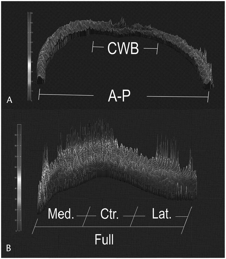
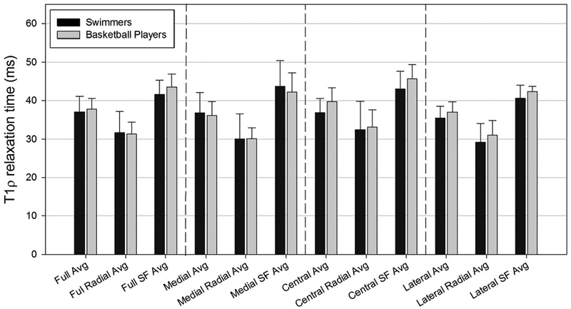
Main outcome measures: Average T1ρ relaxation times (ART) for patellar and femoral cartilage to analyze defined regions and depth and modified International Cartilage Repair Society classification.
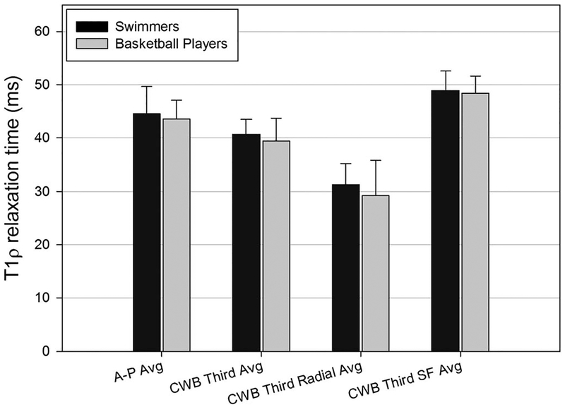
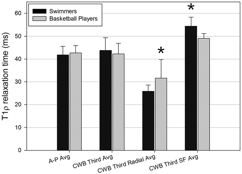
Results: Statistical analyses showed that ART of radial zone of central third weight-bearing region of cartilage in basketball players was significantly greater (P = 0.041) than swimmers and ART of the superficial zone in basketball players was significantly less (P = 0.003) than that of swimmers. For both groups, the ART of superficial zones were significantly greater than that of radial zones (P < 0.001). Four impact athletes showed macroscopic changes (none in nonimpact cohort).
Conclusions: T1ρ MRI detected early changes in articular cartilage of asymptomatic collegiate female impact athletes, with significant differences between cohorts in radial zone of central third weight-bearing region and superficial zones ART. Both cohorts showed increased ART in superficial zone. Four impact athletes showed macroscopic changes.
Clinical relevance: This study demonstrates a quantitative MRI sequence able to detect signal differences in articular cartilage in asymptomatic athletes.
Conflict of interest statement
The authors report no conflicts of interest.
Figures

Similar articles
-
Effects of the Competitive Season and Off-Season on Knee Articular Cartilage in Collegiate Basketball Players Using Quantitative MRI: A Multicenter Study.J Magn Reson Imaging. 2021 Sep;54(3):840-851. doi: 10.1002/jmri.27610. Epub 2021 Mar 24. J Magn Reson Imaging. 2021. PMID: 33763929 Free PMC article.
-
Magnetic Resonance Imaging of Asymptomatic Knees in Collegiate Basketball Players: The Effect of One Season of Play.Clin J Sport Med. 2016 Nov;26(6):483-489. doi: 10.1097/JSM.0000000000000283. Clin J Sport Med. 2016. PMID: 27347867 Free PMC article.
-
Multiparametric MRI characterization of knee articular cartilage and subchondral bone shape in collegiate basketball players.J Orthop Res. 2021 Jul;39(7):1512-1522. doi: 10.1002/jor.24851. Epub 2020 Sep 17. J Orthop Res. 2021. PMID: 32910520 Free PMC article.
-
Advances in magnetic resonance imaging of articular cartilage.J Am Acad Orthop Surg. 2011 Jul;19(7):420-9. doi: 10.5435/00124635-201107000-00005. J Am Acad Orthop Surg. 2011. PMID: 21724921 Review.
-
Advanced MRI Approaches for Evaluating Common Lower Extremity Injuries in Basketball Players: Current and Emerging Techniques.J Magn Reson Imaging. 2024 Jun;59(6):1902-1913. doi: 10.1002/jmri.29019. Epub 2023 Oct 18. J Magn Reson Imaging. 2024. PMID: 37854004 Review.
Cited by
-
Articular Cartilage Reconstruction with Hyaluronate-Based Scaffold Significantly Decreases Pain and Improves Patient's Functioning.J Clin Med. 2023 Nov 27;12(23):7342. doi: 10.3390/jcm12237342. J Clin Med. 2023. PMID: 38068394 Free PMC article.
-
Prevalence of knee osteoarthritis features on magnetic resonance imaging in asymptomatic uninjured adults: a systematic review and meta-analysis.Br J Sports Med. 2019 Oct;53(20):1268-1278. doi: 10.1136/bjsports-2018-099257. Epub 2018 Jun 9. Br J Sports Med. 2019. PMID: 29886437 Free PMC article.
-
T1ρ magnetic resonance imaging quantification of early articular cartilage degeneration in a rabbit model.BMC Musculoskelet Disord. 2015 Nov 19;16:361. doi: 10.1186/s12891-015-0810-0. BMC Musculoskelet Disord. 2015. PMID: 26585246 Free PMC article.
-
Does Cartilage Degenerate in Asymptomatic Hips With Cam Morphology?Clin Orthop Relat Res. 2019 May;477(5):962-971. doi: 10.1097/CORR.0000000000000629. Clin Orthop Relat Res. 2019. PMID: 30801274 Free PMC article.
-
T1ρ magnetic resonance: basic physics principles and applications in knee and intervertebral disc imaging.Quant Imaging Med Surg. 2015 Dec;5(6):858-85. doi: 10.3978/j.issn.2223-4292.2015.12.06. Quant Imaging Med Surg. 2015. PMID: 26807369 Free PMC article. Review.
References
-
- Xia Y Averaged and depth-dependent anisotropy of articular cartilage by microscopic imaging. Semin Arthritis Rheum. 2008;37:317–327. - PubMed
-
- Xia Y Magic-angle effect in magnetic resonance imaging of articular cartilage: a review. Invest Radiol. 2000;35:602–621. - PubMed
-
- Mosher TJ, Dardzinski BJ, Smith MB. Human articular cartilage: influence of aging and early symptomatic degeneration on the spatial variation of T2—preliminary findings at 3 T. Radiology. 2000;214:259–266. - PubMed
Publication types
MeSH terms
Grants and funding
LinkOut - more resources
Full Text Sources
Other Literature Sources
Medical
