
C3 glomerulopathy: consensus report

Matthew C Pickering et al. Kidney Int. 2013 Dec.
Free PMC article

Abstract

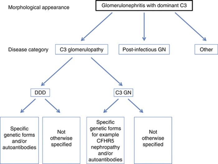
C3 glomerulopathy is a recently introduced pathological entity whose original definition was glomerular pathology characterized by C3 accumulation with absent or scanty immunoglobulin deposition. In August 2012, an invited group of experts (comprising the authors of this document) in renal pathology, nephrology, complement biology, and complement therapeutics met to discuss C3 glomerulopathy in the first C3 Glomerulopathy Meeting. The objectives were to reach a consensus on: the definition of C3 glomerulopathy, appropriate complement investigations that should be performed in these patients, and how complement therapeutics should be explored in the condition. This meeting report represents the current consensus view of the group.


Figures

Figure 1
Figure 1
Immunohistology for C3c. (a) Immunofluorescence in dense deposit disease, (b) immunofluorescence in a case of C3 glomerulonephritis showing predominantly capillary wall staining, and (c) immunoperoxidase in a case of C3 glomerulonephritis showing predominantly mesangial staining.
Figure 2
Figure 2
Electron microscopy in C3 glomerulopathy. (a) Dense deposit disease showing very electron-dense osmiophilic deposit occupying most of the glomerular basement membrane. (b) A case of dense deposit disease in which the highly osmiophilic deposit is only seen in segments of the glomerular basement membrane. (c) A case of C3 glomerulopathy in which there is electron-dense material that is expanding the basement membrane. The material is less electron dense and less well defined than in dense deposit disease. (d) A case of C3 glomerulopathy showing two large subepithelial hump-shaped deposits.
Figure 3
Figure 3
Immunofluorescence in a case of C3 glomerulonephritis illustrating that a small amount of immunoglobulin G (IgG) may be present. (a) C3c and (b) IgG.
Figure 4
Figure 4
A schematic diagram showing an approach to the classification of disease in a biopsy showing the morphological changes of a glomerulonephritis with dominant C3. DDD, dense deposit disease; Post-infectious GN, post-infectious glomerulonephritis.

References

    1. Fakhouri F, Fremeaux-Bacchi V, Noel LH, et al. C3 glomerulopathy: a new classification. Nat Rev Nephrol. 2010;6:494–499. - PubMed
    1. Martinez-Barricarte R, Heurich M, Valdes-Canedo F, et al. Human C3 mutation reveals a mechanism of dense deposit disease pathogenesis and provides insights into complement activation and regulation. J Clin Invest. 2010;120:3702–3712. - PMC - PubMed
    1. Gale DP, de Jorge EG, Cook HT, et al. Identification of a mutation in complement factor H-related protein 5 in patients of Cypriot origin with glomerulonephritis. Lancet. 2010;376:794–801. - PMC - PubMed
    1. Malik TH, Lavin PJ, Goicoechea de Jorge E, et al. A hybrid CFHR3-1 gene causes familial C3 glomerulopathy. J Am Soc Nephrol. 2012;23:1155–1160. - PMC - PubMed
    1. Walker PD, Ferrario F, Joh K, et al. Dense deposit disease is not a membranoproliferative glomerulonephritis. Mod Pathol. 2007;20:605–616. - PubMed

Publication types