. 2013 Nov 21;503(7476):397-401.
doi: 10.1038/nature12655. Epub 2013 Oct 30.

Staphylococcus δ-toxin induces allergic skin disease by activating mast cells

Affiliations

Staphylococcus δ-toxin induces allergic skin disease by activating mast cells

Yuumi Nakamura et al. Nature. .

Abstract

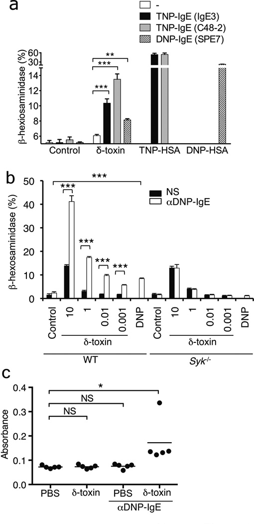
Atopic dermatitis is a chronic inflammatory skin disease that affects 15-30% of children and approximately 5% of adults in industrialized countries. Although the pathogenesis of atopic dermatitis is not fully understood, the disease is mediated by an abnormal immunoglobulin-E immune response in the setting of skin barrier dysfunction. Mast cells contribute to immunoglobulin-E-mediated allergic disorders including atopic dermatitis. Upon activation, mast cells release their membrane-bound cytosolic granules leading to the release of several molecules that are important in the pathogenesis of atopic dermatitis and host defence. More than 90% of patients with atopic dermatitis are colonized with Staphylococcus aureus in the lesional skin whereas most healthy individuals do not harbour the pathogen. Several staphylococcal exotoxins can act as superantigens and/or antigens in models of atopic dermatitis. However, the role of these staphylococcal exotoxins in disease pathogenesis remains unclear. Here we report that culture supernatants of S. aureus contain potent mast-cell degranulation activity. Biochemical analysis identified δ-toxin as the mast cell degranulation-inducing factor produced by S. aureus. Mast cell degranulation induced by δ-toxin depended on phosphoinositide 3-kinase and calcium (Ca(2+)) influx; however, unlike that mediated by immunoglobulin-E crosslinking, it did not require the spleen tyrosine kinase. In addition, immunoglobulin-E enhanced δ-toxin-induced mast cell degranulation in the absence of antigen. Furthermore, S. aureus isolates recovered from patients with atopic dermatitis produced large amounts of δ-toxin. Skin colonization with S. aureus, but not a mutant deficient in δ-toxin, promoted immunoglobulin-E and interleukin-4 production, as well as inflammatory skin disease. Furthermore, enhancement of immunoglobulin-E production and dermatitis by δ-toxin was abrogated in Kit(W-sh/W-sh) mast-cell-deficient mice and restored by mast cell reconstitution. These studies identify δ-toxin as a potent inducer of mast cell degranulation and suggest a mechanistic link between S. aureus colonization and allergic skin disease.


Figures

Figure 1
Figure 1. δ-toxin from S. aureus induces MC degranulation in vitro and in vivo
a, β-Hexosaminidase activity released to the extracellular media of BMCMCs stimulated with medium alone (Control) or indicated stimuli including different concentrations of culture supernatant of S. aureus 8325-4 (S.a sup). b, β-Hexosaminidase activity in supernatants of MC/9 cells stimulated with 10% of culture supernatant from LAC S. aureus wild-type (LAC wt) or isogenic mutants deficient in PSMα peptides (LAC Δpsmα), PSMβ peptides (LAC Δpsmβ), δ-toxin (LAC Δhld), LAC wild-type expressing vector alone (LAC pTXΔ16), LAC deficient in δ-toxin expressing vector alone (LACΔhld pTXΔ16) and strain complemented with δ-toxin plasmid (LACΔhld pTXΔhld). Control represents 10% TSB medium. c, Histamine concentrations in culture supernatant of fetal skin-derived MCs (FSMCs) after stimulation with indicated stimuli including synthetic δ-toxin at 30 µg ml−1 for 15 min. Data represent means ± s.d. of triplicate cultures. Results are representative of at least 3 independent experiments (a-c). P value refers to comparisons between experimental and control groups (a-c). d, Electromicroscopic images of FSMCs stimulated with synthetic δ-toxin (30 µg ml−1) for 15 min. Images of unstimulated (Cont) and ionomycin-treated FSMCs are also shown. Representative of at least 20 images. e, δ-toxin expression in Staphylococcus culture supernatants (0.5 µl per well). Loading of lanes with synthetic δ-toxin (10 ng, 100 ng) is shown as reference. Representative of three experiments. f, C57BL6 (WT) and MC-deficient (Kit w-sh/w-sh) mice were injected intradermally into the left and right ears with δ-toxin (100 µg) or PBS, respectively. One representative mouse for each group is shown. Representative of 8 mice per group. g, Quantification of Evans blue extracted from skin tissue of WT, Kit w-sh/w-sh, Kit w-sh/w-sh reconstituted with BMCMCs is shown. Dots represent individual ear samples from 2 independent experiments. NS; no significant; *P < 0.05; **P < 0.01 ; ***P < 0.001, 2-tailed t test
Figure 2
Figure 2. δ-toxin-induced MC degranulation depends on Ca2+ influx/PI3K pathway, but is independent of Syk
a, FSMCs loaded with the fluorescent Ca2+ indicator Fluo-4AM with or without EGTA were stimulated for 50 sec. Baseline fluorescence (red) was measured, and then the MCs were stimulated with indicated stimuli and fluorescence shift (green) was measured. RFU, relative fluorescence units. b, c, β-Hexosaminidase activity in culture supernatants of FSMCs pretreated with EGTA (b) or LY294002 (c) stimulated with medium alone (Crtl), ionomycin, DNP-HSA (DNP) plus anti DNP-IgE or δ-toxin (10 µg ml−1). d, β-Hexosaminidase activity in culture supernatants of FSMCs derived from Syk−/− and wild-type (WT) mice stimulated with indicated concentration of δ-toxin (µg ml−1). Data represent means ± s.d. of triplicates cultures and representative of at least 3 independent experiments (b-d). NS; no significant; *P < 0.05; **P < 0.01 ; ***P < 0.001, 2-tailed t test
Figure 3
Figure 3. Antigen-independent IgE signaling enhances δ-toxin-induced MC activation
a, β-Hexosaminidase activity in culture supernatants of FSMCs stimulated with or without anti DNP-IgE or TNP-IgE and then re-stimulated with δ-toxin (0.01 µg ml−1), DNP-HSA (DNP) or TNP-HSA (TNP). b, β-Hexosaminidase activity in culture supernatants of FSMCs derived from Syk−/− and wild-type mice (WT) pretreated with or without anti DNP-IgE, and then stimulated with indicated concentration of δ-toxin (µg ml−1). Representative of at least 3 independent experiments. **P < 0.01; ***P < 0.001, 2-tailed t test (a,b). c, Quantification of Evans blue extracted from skin tissue of C57BL6 mice injected intradermally into the left and right ears with δ-toxin (5 µg) or PBS, respectively. Data represent means ± s.d. of triplicate cultures and representative of at least 3 independent experiments (a,b). Dots represent individual ear samples. Representative of 2 independent experiments (c). NS; no significant; *P < 0.05, one-way ANOVA with Tukey post-hoc test for multiple comparisons
Figure 4
Figure 4. Staphyloccocus δ-toxin promotes IgE production and inflammatory skin disease via mast cells
a, S. aureus colonization and OVA sensitization protocol. Mice were colonized epicutaneously with 108 CFU S. aureus using a gauze patch for 1 week. For OVA sensitization, a patch containing OVA or PBS was applied to the same skin site 2 weeks after S. aureus inoculation. b, Skin disease score 1 week post colonization with wild-type and δ-toxin mutant (Δhld) S. aureus or treated with PBS. **P < 0.01; ***P < 0.001, Kruskal-Wallis test with post-hoc Dunn’s test for multiple comparisons. c, Skin phenotype and histopathology of BALB/c mice colonized with S. aureus or treated with PBS. Skin sections were stained with H&E. Bar = 100 µm. Inset shows high power image with neutrophil-rich inflammation. Representative of 14 mice per group. d, Number of inflammatory cells in skin of BALB/c mice colonized with S. aureus or treated with PBS. Results depicted as number of inflammatory cells per high power field (hpf). Error bars represent means ± s. e. m. e, Serum levels of IgE in BALB/c mice colonized with S. aureus or treated with PBS at 1 and 3 weeks post colonization with S. aureus. f, Serum levels of OVA-specific IgE after OVA sensitization in BALB/c mice colonized with S. aureus or treated with PBS. g, Skin disease score in C57BL/6 (B6), MC-deficient (KitW-sh/W-sh) and MC-deficient (KitW-sh/W-sh) mice reconstituted with MCs at 1 week after the inoculation with S. aureus. h, Serum levels of total IgE 1 week after colonization of B6, KitW-sh/W-sh and KitW-sh/W-sh mice reconstituted with MCs with wild-type and δ-toxin mutant (Δhld) S. aureus or treated with PBS. Dots represent individual mice pooled from two independent experiments. *P < 0.05; **P < 0.01 ; ***P < 0.001, one-way ANOVA with Tukey post-hoc test for multiple comparisons (e-h)

References

    1. Williams H, Flohr C. How epidemiology has challenged 3 prevailing concepts about atopic dermatitis. J Allergy Clin Immunol. 2006;118:209–213. - PubMed
    1. Elias PM, Steinhoff M. "Outside-to-inside" (and now back to "outside") pathogenic mechanisms in atopic dermatitis. J Invest Dermatol. 2008;128:1067–1070. - PMC - PubMed
    1. Liu FT, Goodarzi H, Chen HY. IgE, mast cells, and eosinophils in atopic dermatitis. Clin Rev Allergy Immunol. 2011;41:298–310. - PubMed
    1. Galli SJ, Tsai M. IgE and mast cells in allergic disease. Nat Med. 2012;18:693–704. - PMC - PubMed
    1. Rudikoff D, Lebwohl M. Atopic dermatitis. Lancet. 1998;351:1715–1721. - PubMed

Supplementary References

    1. Nilsson IM, Hartford O, Foster T, Tarkowski A. Alpha-toxin and gamma-toxin jointly promote Staphylococcus aureus virulence in murine septic arthritis. Infect Immun. 1999;67:1045–1049. - PMC - PubMed
    1. Stoll H, Dengjel J, Nerz C, Gotz F. Staphylococcus aureus deficient in lipidation of prelipoproteins is attenuated in growth and immune activation. Infect Immun. 2005;73:2411–2423. - PMC - PubMed
    1. Mack D, et al. Characterization of transposon mutants of biofilm-producing Staphylococcus epidermidis impaired in the accumulative phase of biofilm production: genetic identification of a hexosamine-containing polysaccharide intercellular adhesin. Infect Immun. 1994;62:3244–3253. - PMC - PubMed
    1. Francis KP, et al. Monitoring bioluminescent Staphylococcus aureus infections in living mice using a novel luxABCDE construct. Infect Immun. 2000;68:3594–3600. - PMC - PubMed
    1. Bae T, Schneewind O. Allelic replacement in Staphylococcus aureus with inducible counter-selection. Plasmid. 2006;55:58–63. - PubMed

Publication types

MeSH terms