. 2014 Jan;15(1):25-32.
doi: 10.1038/gene.2013.57. Epub 2013 Oct 31.

Differential CTLA-4 expression in human CD4+ versus CD8+ T cells is associated with increased NFAT1 and inhibition of CD4+ proliferation

Affiliations

Differential CTLA-4 expression in human CD4+ versus CD8+ T cells is associated with increased NFAT1 and inhibition of CD4+ proliferation

D V Chan et al. Genes Immun. 2014 Jan.

Abstract

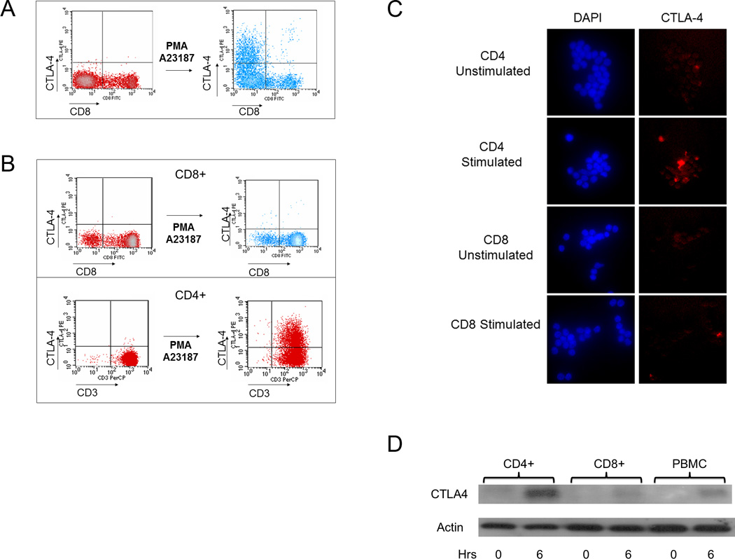
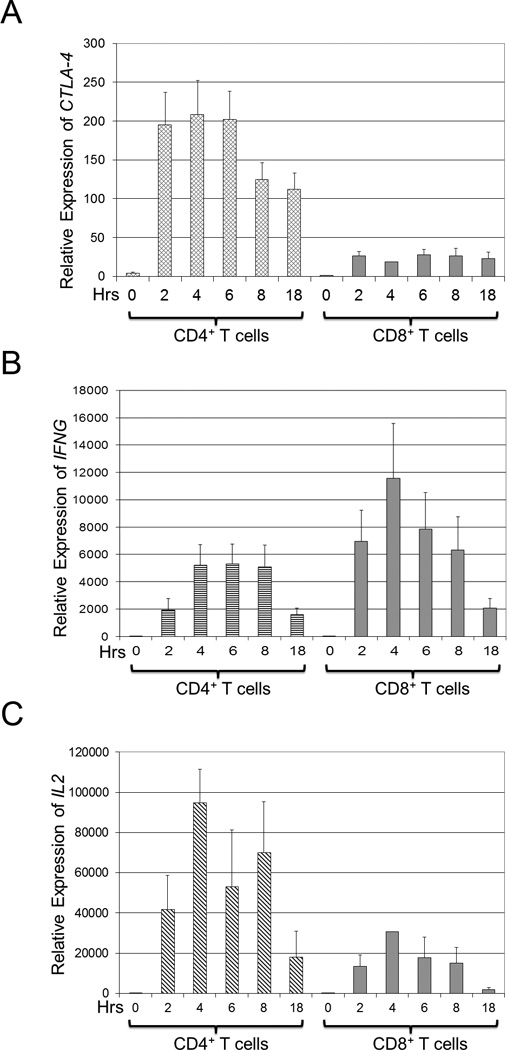
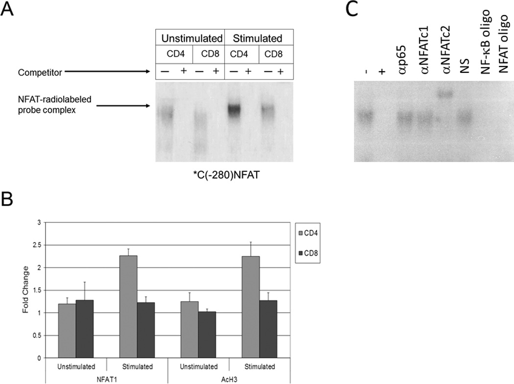
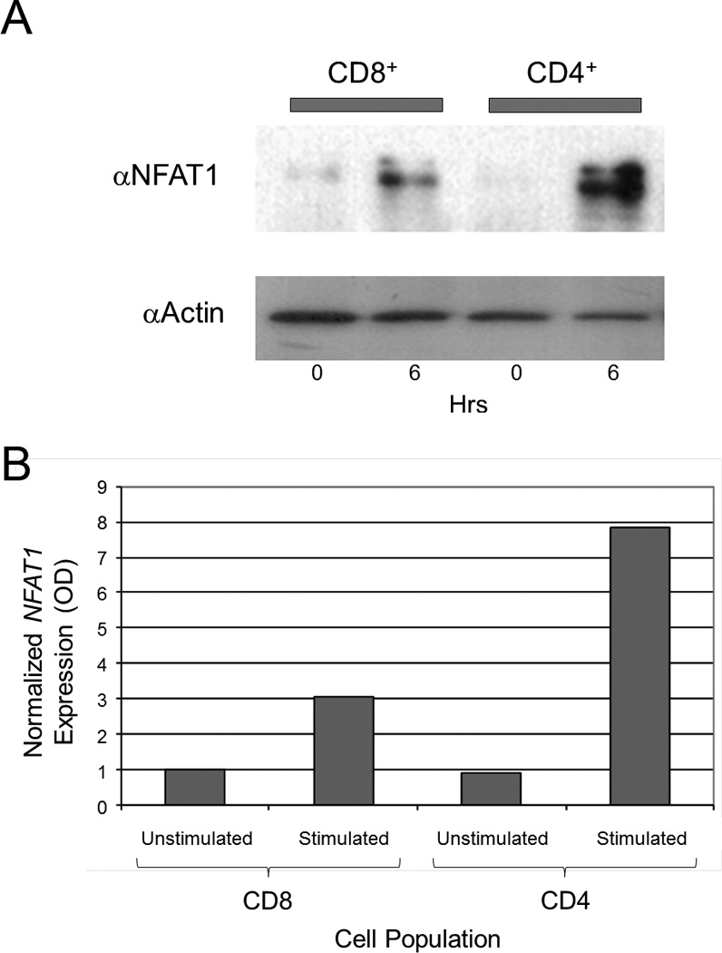
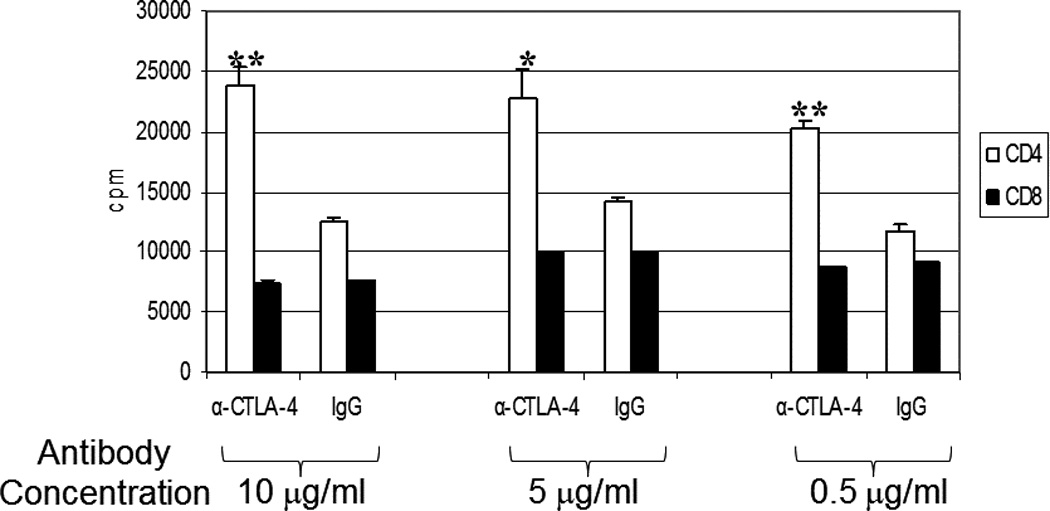
Cytotoxic T-lymphocyte-associated protein 4 (CTLA-4) is a costimulatory molecule that negatively regulates T-cell activation. Originally identified in murine CD8(+) T cells, it has been found to be rapidly induced on human T cells. Furthermore, CTLA-4 is expressed on regulatory T cells. Clinically, targeting CTLA-4 has clinical utility in the treatment of melanoma. Whether the expression of CTLA-4 is differentially regulated in CD8(+) vs CD4(+) human T cells is unclear. Here, we analyzed CTLA-4 in normal human CD4(+) and CD8(+) T-cell subsets and show for the first time that CTLA-4 is expressed significantly higher in the CD4(+) T cells than in CD8(+) T cells. CTLA-4 is higher at the protein and the transcriptional levels in CD4(+) T cells. This increase is due to the activation of the CTLA-4 promoter, which undergoes acetylation at the proximal promoter. Furthermore, we show that blocking CTLA-4 on CD4(+) T cells permits greater proliferation in CD4(+) vs CD8(+) cells. These findings demonstrate a differential regulation of CTLA-4 on CD4(+) and CD8(+) T-cell subsets, which is likely important to the clinical efficacy for anti-CTLA-4 therapies. The findings hint to strategies to modulate CTLA-4 expression by targeting epigenetic transcription to alter the immune response.


Conflict of interest statement

Conflict of Interest Disclosure

None of the authors have any competing financial interests to declare in relation to the work described in this manuscript.

Figures

Figure 1
Figure 1. CTLA-4 is preferentially induced in CD4 vs. CD8 T cells
(a) The level of CTLA-4 expression was measured by flow cytometry before and after stimulation with PMA/A23187 as described in the Materials and Methods. Few CD8+ T cells express CTLA-4, suggesting that CTLA-4 is expressed mainly in non-CD8+ T cells. The results are representative of findings from three normal volunteers. (b) CD4 and CD8 T cells were purified using negative selection as described in the Materials and Methods. After stimulation with PMA/A23187, CTLA-4 expression was measured. CTLA-4 was minimal on the purified CD8+ subset (top panel) but was detected on the purified CD4+ subset (bottom panel). The results in each panel are representative of findings from three normal volunteers. (c) CTLA-4 is preferentially increased in stimulated CD4+ vs. CD8+ cells as assessed by immunofluorescence. Purified cells were stimulated with PMA/A23187 and fixed as described. Cells were stained with DAPI and anti-CTLA-4-PE as described in the Materials and Methods. Results are representative of three independent experiments. d) CD4+ and CD8+ T cells as well as bulk PBMCs were analyzed for total CTLA-4 expression. 10 million of each purified cell type was used in conditions following stimulation by PMA/A23187 as indicated. Following treatment with PMA/A23187 for 0 or 6 hours (horizontal axis), total protein was isolated as described in the Materials and Methods. 10 ug of total protein was separated by SDS-PAGE, transferred to PVDF membrane and immunoblotted for total CTLA-4 levels using anti-CTLA-4 followed by visualization by chemiluminescence.
Figure 2
Figure 2. CTLA-4 gene expression is preferentially increased in CD4 T cells
CD4+ and CD8+ T cells were isolated by negative selection from PBMCs as described in the Materials and Methods. Equal numbers of purified cells were stimulated with PMA/A23187 for 0, 2, 4, 6, 8, or 18 hours (hrs, horizontal axis), and total RNA was isolated and analyzed by qRT-PCR as described above. Results are depicted as average values ± s.d. of three normal volunteers. (a) The relative expression of CTLA-4 is higher in CD4+ compared to CD8+ T cells. CTLA-4 gene expression was normalized to CTLA-4 gene expression in unstimulated CD4+ T cells. (b) The relative expression of IFN-γ (IFNG) is higher in CD8+ compared to CD4+ T cells. IFNG gene expression was normalized to IFNG gene expression in unstimulated CD4+ T cells. (c) The relative expression of IL-2 (IL2) is higher in CD4+ compared to CD8+ T cells. IL2 gene expression was normalized to IL-2 gene expression in unstimulated CD4+ T cells.
Figure 3
Figure 3. Increased NFAT activity is found in CD4+ vs. CD8+ T cells at the CTLA-4 promoter
a) Protein extracts from unstimulated and stimulated CD4+ and CD8+ T cells were incubated with a radiolabelled probe representing an NFAT binding site in the CTLA-4 promoter (*C(−280)NFAT). Subsequently, mixtures were then incubated with or without excess competitor unlabeled probe (+ or −) and NFAT1 binding was assayed via 4% polyacrylamide gel electrophoresis. b) CHIP assays were performed on the CTLA-4 promoter region using anti-NFAT1 (left panel) and anti-acetylated histone H3 (AcH3, right panel) antibodies in unstimulated and stimulated CD4+ and CD8+ T cells. Normalization for equal cell numbers was 10% of the input DNA. Results in each panel are averages from two independent experiments. c) Gel shift mobility assay (EMSA) was performed as described above with our without excess competitor unlabeled probe (+ or −) and gel shift was assessed using antibodies to the p65 subunit of NF-κB, NFAT1, and NFAT2. Normal serum (NS), NF-κb oligonucleotides (NF-κb oligo) and NFAT oligonucleotides (NFAT oligo) were used as additional controls.
Figure 4
Figure 4. NFAT1 is increased in CD4+ T cells compared to CD8+ T cells
(a) NFAT1 is increased in stimulated CD4+ cells vs. stimulated CD8+ cells by immunoblot. Total protein was isolated from purified unstimulated (0 hours, horizontal axis) and stimulated (6 hours, horizontal axis) CD4+ and CD8+ T cells. Equal amount amounts of protein (10 µg) was analyzed by SDS-PAGE and immunoblotted against NFAT1 with an anti-NFAT1 antibody (αNFATc2) as shown. The level of actin was determined by immunoblotting the same membrane with an anti-actin antibody (αActin) after stripping the anti-NFAT1 to control for equal protein loading. Results are representative of three independent experiments. (b) NFAT1 expression was measured by qRT-PCR and normalized to β2-microglobulin. Stimulated CD4+ cells show higher relative expression of NFAT1 compared to stimulated CD8+ cells.
Figure 5
Figure 5. CTLA-4 expression on CD4+ T cells reduces the proliferative potential of CD4+ T cells
Purified CD4+ or CD8+ T cells were cultured as described in the Materials and Methods. Mixed lymphocyte reaction was performed in the presence of anti-CTLA-4 or IgG isotype control antibody was titrated at 10 µg/ml, 5 µg/ml, or 0.5 µg/ml with CD4+ or CD8+ T cells as indicated over 7 days followed by additional of H3-thymidine. Average cpm were calculated ± s.d. p values were calculated comparing the difference in cpm proliferation in CD4+ vs. CD8+ T cells and were < 0.01 (*) or < 0.001 (**) as indicated. Results are representative of 2 independent experiments.

References

    1. Dariavach P, Mattéi M-G, Golstein P, Lefranc M-P. Human Ig superfamily CTLA-4 gene: chromosomal localization and identity of protein sequence between murine and human CTLA-4 cytoplasmic domains. European Journal of Immunology. 1988;18(12):1901–1905. - PubMed
    1. Harper K, Balzano C, Rouvier E, Mattei M, Luciani M, Golstein P. CTLA-4 and CD28 activated lymphocyte molecules are closely related in both mouse and human as to sequence, message expression, gene structure, and chromosomal location. The Journal of Immunology. 1991;147(3):1037–1044. - PubMed
    1. Auchincloss H, Turka LA. CTLA-4: not all costimulation is stimulatory. J Immunol. 2011;187(7):3457–3458. - PMC - PubMed
    1. Lindsten T, Lee K, Harris E, Petryniak B, Craighead N, Reynolds P, et al. Characterization of CTLA-4 structure and expression on human T cells. The Journal of Immunology. 1993;151(7):3489–3499. - PubMed
    1. Friedline RH, Brown DS, Nguyen H, Kornfeld H, Lee J, Zhang Y, et al. CD4+ regulatory T cells require CTLA-4 for the maintenance of systemic tolerance. J Exp Med. 2009;206(2):421–434. - PMC - PubMed

Publication types