

Dermoscopic features of thin melanomas: a comparative study of melanoma in situ and invasive melanomas smaller than or equal to 1mm
- PMID: 24173175
- PMCID: PMC3798346
- DOI: 10.1590/abd1806-4841.20132017
Dermoscopic features of thin melanomas: a comparative study of melanoma in situ and invasive melanomas smaller than or equal to 1mm
Abstract
Background: Dermoscopy allows the early detection of melanomas. The preoperative determination of Breslow index by dermoscopy could be useful in planning the surgical approach and in selecting patients for sentinel lymph node biopsy.
Objectives: This study aims at describing the dermoscopic features of thin melanomas and comparing melanomas in situ with invasive melanomas less than or equal to 1 mm thick.
Methods: This was an observational retrospective study in which the dermoscopy photographs of 41 thin melanomas were evaluated. Three observers evaluated together 14 dermoscopic criteria.
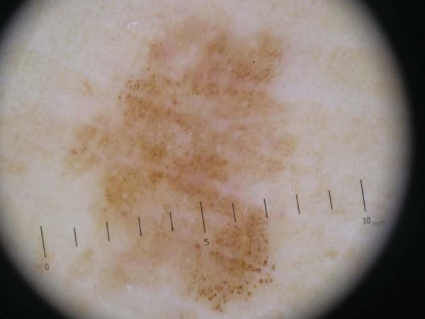
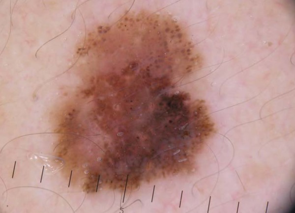
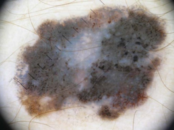
Results: Among thin melanomas, the most frequent criteria were presence of asymmetry in two axes in 95% of cases (39 cases), 3 or more colors in 80.4% of cases (33 cases), atypical dots or globules in 58.5% of cases (24 cases) and atypical network or streaks in 53.6% of cases (22 cases). The group of invasive melanomas presented with a higher frequency and statistical significance (p <0.05) 3 or more colors (OR: 16.1), milky red areas (OR: 4.8) and blue-white veil (OR: 20.4), and a greater tendency to have streaks or atypical network (OR: 3.66).
Conclusions: Thin melanomas tend to have asymmetry in the two axes, 3 or more colors, atypical dots or globules and atypical network or streaks. Melanomas in situ tend to have up to 2 colors, no blue-white veil and no milky red area. Invasive melanomas tend to have 3 or more colors, a milky red area, blue-white veil, and atypical network or streaks. Further studies are needed to confirm these findings.
FUNDAMENTOS: A dermatoscopia propicia o diagnóstico mais precoce do melanoma. A estimativa préoperatória da espessura de Breslow através da dermatoscopia poderia ser útil no planejamento da conduta cirúrgica e seleção dos pacientes para a biópsia de linfonodo sentinela.
OBJETIVOS: Este estudo objetiva descrever as características dermatoscópicas encontradas em melanomas finos e comparar melanomas in situ com melanomas invasivos menores ou iguais a 1 mm.
MÉTODOS: Trata-se de estudo observacional, retrospectivo, no qual avaliouse o registro fotográfico da dermatoscopia de 41 melanomas finos. Três observadores avaliaram em conjunto 14 critérios dermatoscópicos.
RESULTADOS: Dentre os melanomas finos, os critérios mais encontrados foram: presença de assimetria nos dois eixos em 95% (39 casos), 3 ou mais cores em 80,4% (33 casos), pontos ou glóbulos atípicos em 58,5% (24 casos) e rede atípica ou estrias radiadas em 53,6% (22 casos). O grupo dos melanomas invasivos apresentou com maior frequência e significância estatística (p<0,05) a presença de 3 ou mais cores (OR: 16,1), áreas vermelho-leitosas (OR: 4,8) e véu (OR: 20,4), além de uma maior tendência em apresentar rede atípica ou estrias radiadas (OR: 3,66).
CONCLUSÕES: Os melanomas finos tendem a apresentar assimetria nos dois eixos, 3 ou mais cores, pontos ou glóbulos atípicos e rede atípica ou estrias radiadas. Os melanomas in situ tendem a apresentar até 2 cores, ausência de véu e de área vermelho-leitosa. Os melanomas invasivos tendem a exibir 3 cores ou mais, área vermelho-leitosa, véu, rede atípica ou estrias radiadas. Outros estudos são necessários para a confirmação dos achados.
Conflict of interest statement
Conflict of Interest: None
Figures
Similar articles
-
Analysis of the dermoscopic features of excised melanomas and their relation with tumor thickness in a tertiary hospital in Brazil.Int J Dermatol. 2024 Aug;63(8):1064-1070. doi: 10.1111/ijd.17047. Epub 2024 Feb 28. Int J Dermatol. 2024. PMID: 38415856
-
Dermoscopic findings for the early detection of melanoma: an analysis of 200 cases.Actas Dermosifiliogr. 2014 Sep;105(7):683-93. doi: 10.1016/j.ad.2014.01.008. Epub 2014 Apr 3. Actas Dermosifiliogr. 2014. PMID: 24704190 English, Spanish.
-
Amelanotic/hypomelanotic melanoma: clinical and dermoscopic features.Br J Dermatol. 2004 Jun;150(6):1117-24. doi: 10.1111/j.1365-2133.2004.05928.x. Br J Dermatol. 2004. PMID: 15214897
-
Assessment of Diagnostic Accuracy of Dermoscopic Structures and Patterns Used in Melanoma Detection: A Systematic Review and Meta-analysis.JAMA Dermatol. 2021 Sep 1;157(9):1078-1088. doi: 10.1001/jamadermatol.2021.2845. JAMA Dermatol. 2021. PMID: 34347005 Free PMC article.
-
Role of In Vivo Reflectance Confocal Microscopy in the Analysis of Melanocytic Lesions.Acta Dermatovenerol Croat. 2018 Apr;26(1):64-67. Acta Dermatovenerol Croat. 2018. PMID: 29782304 Review.
Cited by
-
Dermoscopic evaluation of superficial spreading melanoma.An Bras Dermatol. 2021 Mar-Apr;96(2):139-147. doi: 10.1016/j.abd.2020.06.012. Epub 2021 Feb 1. An Bras Dermatol. 2021. PMID: 33637398 Free PMC article.
-
Discrimination Between Invasive and In Situ Melanomas Using Clinical Close-Up Images and a De Novo Convolutional Neural Network.Front Med (Lausanne). 2021 Sep 14;8:723914. doi: 10.3389/fmed.2021.723914. eCollection 2021. Front Med (Lausanne). 2021. PMID: 34595193 Free PMC article.
-
Dermoscopy of external ear melanoma (EEM).Arch Dermatol Res. 2023 Jul;315(5):1381-1387. doi: 10.1007/s00403-022-02342-2. Epub 2022 Mar 17. Arch Dermatol Res. 2023. PMID: 35298674
-
Assessment of melanoma thickness based on dermoscopy images: an open, web-based, international, diagnostic study.J Eur Acad Dermatol Venereol. 2022 Nov;36(11):2002-2007. doi: 10.1111/jdv.18436. Epub 2022 Jul 26. J Eur Acad Dermatol Venereol. 2022. PMID: 35841304 Free PMC article.
-
The Importance of Dermoscopy in Early Recognition of Melanoma in Situ.Curr Health Sci J. 2019 Oct-Dec;45(4):366-371. doi: 10.12865/CHSJ.45.04.04. Epub 2019 Dec 30. Curr Health Sci J. 2019. PMID: 32110438 Free PMC article.
References
-
- Brasil. Ministério da Saúde. Instituto Nacional de Câncer José Alencar Gomes da Silva (INCA) Estimativa 2012: incidência de câncer no Brasil. Rio de Janeiro: INCA; 2011. [acesso 24 Jun 2011]. pp. 49–50. Disponível em: http://www.inca.gov.br/estimativa/2012/index.asp?ID=1.
-
- Bafounta ML, Beauchet A, Aegerter P, Saiag P. Is dermoscopy (epiluminescence microscopy) useful for the diagnosis of melanoma? Results of a meta-analysis using techniques adapted to the evaluation of diagnostic tests. Arch Dermatol. 2001;137:1343–1350. - PubMed
-
- Mayer J. Systematic review of the diagnostic accuracy of dermatoscopy in detecting malignant melanoma. Med J Aust. 1997;167:206–210. - PubMed
-
- Argenziano G, Cerroni L, Zalaudek I, Staibano S, Hofmann-Wellenhof R, Arpaia N, et al. Accuracy in melanoma detection: A 10-year multicenter survey. J Am Acad Dermatol. 2012;67:54–59. - PubMed
Publication types
MeSH terms
LinkOut - more resources
Full Text Sources
Other Literature Sources
Medical