. 2014 Jan;88(1):503-15.
doi: 10.1128/JVI.01373-13. Epub 2013 Oct 30.

A novel cytotoxic sequence contributes to influenza A viral protein PB1-F2 pathogenicity and predisposition to secondary bacterial infection

Affiliations

A novel cytotoxic sequence contributes to influenza A viral protein PB1-F2 pathogenicity and predisposition to secondary bacterial infection

Irina V Alymova et al. J Virol. 2014 Jan.

Abstract

Enhancement of cell death is a distinguishing feature of H1N1 influenza virus A/Puerto Rico/8/34 protein PB1-F2. Comparing the sequences (amino acids [aa] 61 to 87 using PB1-F2 amino acid numbering) of the PB1-F2-derived C-terminal peptides from influenza A viruses inducing high or low levels of cell death, we identified a unique I68, L69, and V70 motif in A/Puerto Rico/8/34 PB1-F2 responsible for promotion of the peptide's cytotoxicity and permeabilization of the mitochondrial membrane. When administered to mice, a 27-mer PB1-F2-derived C-terminal peptide with this amino acid motif caused significantly greater weight loss and pulmonary inflammation than the peptide without it (due to I68T, L69Q, and V70G mutations). Similar to the wild-type peptide, A/Puerto Rico/8/34 elicited significantly higher levels of macrophages, neutrophils, and cytokines in the bronchoalveolar lavage fluid of mice than its mutant counterpart 7 days after infection. Additionally, infection of mice with A/Puerto Rico/8/34 significantly enhanced the levels of morphologically transformed epithelial and immune mononuclear cells recruited in the airways compared with the mutant virus. In the mouse bacterial superinfection model, both peptide and virus with the I68, L69, and V70 sequence accelerated development of pneumococcal pneumonia, as reflected by increased levels of viral and bacterial lung titers and by greater mortality. Here we provide evidence suggesting that the newly identified cytotoxic sequence I68, L69, and V70 of A/Puerto Rico/8/34 PB1-F2 contributes to the pathogenesis of both primary viral and secondary bacterial infections.


Figures

FIG 1
FIG 1
Amino acid sequences of influenza A virus PB1-F2 proteins. Sequences are shown for selected influenza A virus strains. Green shading highlights amino acids that we propose are a cause of a high level of cell death due to the A/Puerto Rico/8/1934 (H1N1) PB1-F2 protein.
FIG 2
FIG 2
Amino acid sequence I68, L69, and V70 of PR8 PB1-F2 enhances death in various cell lines. (A) Exposure to 27-mer peptides derived from the C-terminal region of the PR8 or Wuh95 PB1-F2 proteins (see Fig. 1 for definition; final concentration for each peptide, 100 μM) or PBS for 1 h. The PR8/C and Wuh95/C + 3 PB1-F2-derived peptides contained the I68, L69, and V70 sequence. In Wuh95/C + 3, this sequence was introduced by mutations T68I, Q69L, and G70V. Wuh95/C and PR8/C-3 did not have the I68, L69, and V70 motif. In PR8/C-3, this motif was knocked out by mutations I68T, L69Q, and V70G. The proportions of live and dead cells in the samples were determined by exclusion of trypan blue. An asterisk indicates a significant (P < 0.05) difference by ANOVA compared with the results for the PBS or peptides without the I68, L69, and V70 sequence (PR8/C-3 and Wun95/C). (B) Image of live (unstained) and dead (stained with Sytox green nucleic acid stain) MDCK cells exposed to 80 μM PR8 or Wuh95 PB1-F2-derived peptides for 30 min. (C) Intracellular distribution of PR8 PB1-F2-derived peptides. Representative confocal Z-stack images of MDCK cells exposed to 50 μM (final concentration) PR8/C and PR8/C-3 biotinylated peptides for 1 h and stained with streptavidin:Cy2 are shown. Green staining represents PB1-F2-derived peptide, and blue represents nuclear staining.
FIG 3
FIG 3
MOMP promotion by the I68, L69, and V70 sequence of PR8 PB1-F2. Western blot analysis of cytochrome c release into the supernatant after exposure of heavy membrane fractions (mitochondria) from mouse liver to 1,000 μM PR8/C (with the I68, L69, and V70 sequence) or PR8/C-3 (with I68T, L69Q, and V70G mutations) or PR8/C peptide with single substitutions at the desired residues for 45 min at 37°C was performed. The pellet fraction contains intact mitochondria with unreleased cytochrome c. C8-BID was used as a positive control for mammalian mitochondria. Arrows show cytochrome c in supernatant or pellet fractions.
FIG 4
FIG 4
Growth kinetics of PR8 and PR8-3 in various cell lines. MDCK, A549, or U937 cells were infected with PR8 or an isogenic mutant altered at I68T, L69Q, and V70G of the PB1-F2 of PR8 (PR8-3) at MOI = 0.01 (A) or 3.0 (B). Virus titers were determined for culture supernatant fluids at the times indicated on the x axis. Error bars indicate the standard deviations (SD) of the means of the results from three independent experiments.
FIG 5
FIG 5
Expression and distribution of PB1-F2 proteins from PR8 and PR8-3. MDCK cell monolayers were infected with PR8 or PR8-3 virus at MOI = 3.0 for 8 h. The PR8-3 had I68T, L69Q, and V70G mutations in the PB1-F2 amino acid sequence. (A) Using confocal microscopy and antibodies specific for viral PB1-F2 or cell mitochondria, the sum binary fluorescent intensities were determined for five fields containing ∼1,000 cells total. Cell nuclei were visualized with DAPI. Differences in PB1-F2 protein expression (green fluorescent signal) between PR8 and PR8-3 are visually notable in representative fields for each virus. Localization of PB1-F2 to mitochondria is indicated by the yellow color in the merged pictures. (B) Total fluorescent intensities from PB1-F2 proteins from PR8 and PR8-3 are expressed as means ± SD. An asterisk indicates a significant difference (P < 0.05) by ANOVA compared with the PR8 virus. (C) Predominant mitochondrial localization of PB1-F2 proteins from PR8 and PR8-3.
FIG 6
FIG 6
The I68, L69, and V70 sequence of PR8 PB1-F2 increases peptide and virus pathogenicity in mice. (A and B) Weight loss (A) and inflammatory and cytokine responses (B) in BALB/c mice exposed to 15 mg PR8/C- or C-3-derived peptides or PBS (as a control) 3 days after exposure. HPF, high-power field. (C and D) Virus lung titers (C) and inflammatory and cytokine responses (D) in mice infected with 15 PFU per mouse of PR8 and PR8-3. PR8/C-3 peptide and PR8-3 virus had I68T, L69Q, and V70G alterations in the PB1-F2 amino acid sequence. (A) Mice (n = 10) were monitored individually for weight loss; results are presented as mean percentages of starting weight ± SD. (B and D) Total white blood cells (WBCs), neutrophils, macrophages, IgA, and cytokines recovered from bronchoalveolar lavage (BAL) fluid of mice (n = 5) 3 days after peptide exposure (B) or 7 days after viral infection (D). Means ± SD are shown. (C) Growth kinetics of PR8 and PR8-3 viruses in lungs of mice (n = 3 to 5 per time point). An asterisk indicates a significant difference (P < 0.05) by ANOVA (A, B, and D) or by Student's t test (C) compared with the results for the mice exposed to the PR8/C peptide (A and B) or infected with the PR8 virus (C and D).
FIG 7
FIG 7
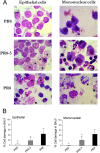
The I68, L69, and V70 sequence of PR8 PB1-F2 induces morphological changes in epithelial and mononuclear cells. Groups of mice were infected with 15 PFU per mouse of PR8 and PR8-3 (whose PB1-F2 had I68T, L69Q, and V70G amino acid alterations) or PBS (as a control) and euthanized 7 days after viral infection. Bronchoalveolar lavage fluid (BALF) of mice (n = 5) was harvested and separated from cells by centrifugation. (A) Epithelial or mononuclear cells from mice given PBS or infected with PR8-3 or PR8 were cytospun, differentially stained, and counted at ×1,000 magnification using light microscopy. Differentiation of morphologically altered (see definition in Materials and Methods) epithelial or mononuclear cells was done using standard morphometric techniques. An asterisk specifies fragmented nuclei; the arrow shows separated cilia or vacuoles in cytoplasm. (B) Cells (at least 1,000) in five random fields per sample were counted to estimate the percentages (means ± SD) of morphologically changed epithelial or mononuclear cells. Asterisks indicate a significant (P < 0.05) difference by ANOVA compared with the results for the PR8-infected group.
FIG 8
FIG 8
The I68, L69, and V70 sequence of PR8 PB1-F2 peptide accelerates bacterial pneumonia in mice. (A to D) Weight loss (A), survival (B), bacterial lung titers (C), and inflammatory cell recruitment (D) in mice exposed to PR8 PB1-F2-derived peptides. BALB/c mice (n = 10 each for panels A, B, and C) were exposed to 10 mg PR8/C or PR8/C-3 peptides or PBS and then challenged 1 day later with a dose of 2,000 CFU of S. pneumoniae (SPn) per mouse. (C) Lungs were collected 1 day after the challenge and titrated for bacterial load. (D) Total white blood cells (WBCs), neutrophils, macrophages, and lymphocytes recovered from the BAL fluid of mice (n = 5) 3 days after bacterial challenge. (A and D) Error bars indicate the SD of the mean. Asterisks indicate a significant difference (P < 0.05) compared with the PR8/C + SPn group by ANOVA (A and D), by Student's t test (C), and by the log-rank test on the Kaplan Meier survival data (B).
FIG 9
FIG 9
Histopathologic changes in the lungs of mice exposed to PR8 PB1-F2-derived peptides in a bacterial pneumonia model. Mice exposed to 10 mg PR8/C or PR8/C-3 (a version of PR8/C altered at I68T, L69Q, and V70G) peptides or PBS were challenged 1 day later with a dose of 2,000 CFU of S. pneumoniae (SPn) per mouse. Sections of lungs stained with hematoxylin and eosin (upper panel) or podoplanin (lower panel [brown staining]) are pictured at ×20 magnification. A semiquantitative grading system was used to assess the degree of injury in each set of lungs. The total scores for all pulmonary lesions (means ± SD) determined for 3 mice per group are shown. Asterisks indicate a significant (P < 0.05) difference by ANOVA compared with the results for the PR8/C + SPn group.
FIG 10
FIG 10
Secondary bacterial pneumonia in PR8- or PR8-3-infected mice. BALB/c mice were infected with a sublethal dose of 15 PFU per mouse of PR8, PR8-3, or PBS and then challenged 7 days later with a sublethal dose of 100 CFU of S. pneumoniae (SPn) per mouse. (A and B) Lungs from 5 to 12 mice were collected 1, 2, and 3 days after bacterial challenge and titrated for viral and bacterial load. (C) Survival of a group of mice (n = 10) was monitored for 21 days after bacterial challenge. Asterisks indicate a significant (P < 0.05) difference compared with the PR8-infected group by Student's t test (A and B) and the log-rank test performed on the Kaplan Meier survival data (C).

References

    1. Wright PF, Neumann G, Kawaoka Y. 2007. Orthomyxoviruses, p 1691–1740 In Knipe DM, Howley PM, Griffin DE, Lamb RA, Martin MA, Roizman B, Straus SE. (ed), Fields virology, 5th ed. Lippincott Williams & Wilkins, Philadelphia, PA
    1. McCullers JA, Huber VC. 2012. Correlates of vaccine protection from influenza and its complications. Hum. Vaccin. Immunother. 8:34–44. 10.4161/hv.8.1.18214 - DOI - PubMed
    1. Chen W, Calvo PA, Malide D, Gibbs J, Schubert U, Bacik I, Basta S, O'Neill R, Schickli J, Palese P, Henklein P, Bennink JR, Yewdell JW. 2001. A novel influenza A virus mitochondrial protein that induces cell death. Nat. Med. 7:1306–1312. 10.1038/nm1201-1306 - DOI - PubMed
    1. Conenello GM, Zamarin D, Perrone LA, Tumpey T, Palese P. 2007. A single mutation in the PB1-F2 of H5N1 (HK/97) and 1918 influenza A viruses contributes to increased virulence. PLoS Pathog. 3:e421. 10.1371/journal.ppat.0030141 - DOI - PMC - PubMed
    1. Conenello GM, Tisoncik JR, Rosenzweig E, Varga ZT, Palese P, Katze MG. 2011. A single N66S mutation in the PB1-F2 protein of influenza A virus increases virulence by inhibiting the early interferon response in vivo. J. Virol. 85:652–662. 10.1128/JVI.01987-10 - DOI - PMC - PubMed

Publication types

Substances