

doi: 10.1128/JVI.02589-13.
Epub 2013 Oct 30.
Immunosuppression facilitates the reactivation of latent papillomavirus infections
Affiliations
- PMID: 24173230
- PMCID: PMC3911712
- DOI: 10.1128/JVI.02589-13
Item in Clipboard
Immunosuppression facilitates the reactivation of latent papillomavirus infections
J Virol.
2014 Jan.
Abstract
At mucosal sites, papillomavirus genomes can persist in the epithelial basal layer following immune-mediated regression. Subsequent T-cell depletion stimulates a 3- to 5-log increase in the viral copy number, to levels associated with productive infection. Reappearance of microlesions was rare within the short time frame of our experiments but was observed in one instance. Our studies provide direct evidence that immunosuppression can trigger the reactivation of latent papillomavirus genomes, as previously proposed in humans.
Figures
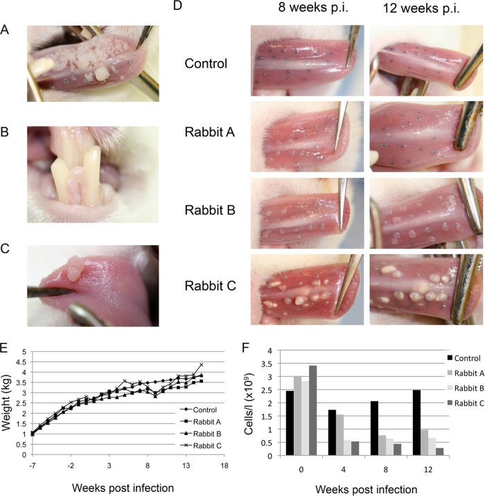
(A) Presence of florid ROPV-induced warts in immunosuppressed rabbits at 15 weeks postinfection. In immunocompetent rabbits, lesions were not detectable beyond 10 weeks. (B and C) In the immunosuppressed rabbits, secondary infections were also apparent at nonscarified sites by 15 weeks postinfection (p.i.). (D) Lesion size and abundance varied among immunosuppressed rabbits (rabbits A, B, and C), depending on the extent of immunosuppression (see panel F). Immunocompetent rabbits developed much smaller lesions (control), with tattoo marks indicating the sites of previous infection by week 12. (E) The cyclosporine-dexamethasone immunosuppression regimen was found not to have a major effect on weight gain in juvenile (growing) rabbits during the time course of the immunosuppression experiment. (F) To establish the extent of immunosuppression, T cell counts of the three immunosuppressed rabbits shown in panel D are shown as gray columns (rabbits A, B, and C); T cell counts of a typical immunocompetent animal are shown as black columns.
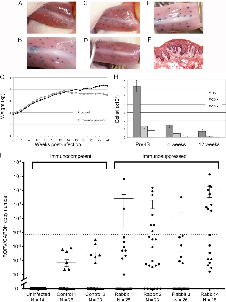
(A to D) Typical appearance of tattoo-marked tongue sites in two ROPV-infected rabbits at 4 weeks postinfection (A and C) and 12 weeks postinfection (B and D). At 4 weeks postinfection (A and C), papillomas are apparent overlying tattoo-marked infection sites. At 12 weeks postinfection in the absence of immunosuppression (B and D), only tattoo marks remain at previous sites of infection. (E) The appearance of tattoo marks in the uninfected control rabbit in which no papilloma lesions developed during the course of the study are indistinguishable from the regressed sites shown in panels B and D. (F) Histology of a single microlesion (with typical papilloma features) detected in rabbit 2 at 12 weeks postimmunosuppression. (G) In adult rabbits, the cyclosporine-dexamethasone immunosuppression regimen generally caused weight loss, with rabbits being culled when their weight loss exceeded 20% of their total body weight. The graph shown is typical of what was seen in repeated experiments. (H) Decline in total lymphocyte counts (TLC; dark gray) and CD4- and CD8-positive (light gray/white) lymphocyte levels following the onset of immunosuppression (IS) in the adult infected rabbits shown in panel I. (I) Numbers of ROPV copies per copy of GAPDH DNA at sites of previous infection in postregression control rabbits (triangles) and postregression immunosuppressed rabbits (circles). Cyclosporine-dexamethasone was administered at week 12 to the immunosuppressed animals, and viral copy numbers were measured at individual tattoo-marked sites at week 24. ROPV/GAPDH copy numbers below the dotted line are those that are typically observed in rabbits with a latent infection or where no ROPV is detectable (12). Elevated copy numbers were seen only at tattoo-marked sites in the immunosuppressed animals.
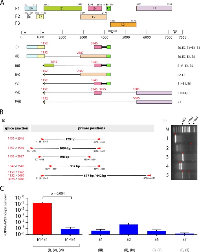
(A) ROPV transcript map based on the analysis of cDNA prepared from a productive rabbit papilloma by a previously described methodology (26). The positions of splice donor and acceptor sites were determined experimentally and are shown in red. Individual transcripts along with their coding capacities are numbered i to vii at the bottom. The positions of the open reading frames and predicted promoter and polyadenylation sites have been reported previously (27) and are marked at the top. (B) Primers used for PCR amplification of ROPV cDNA fragments are indicated by red arrows, and the observed sizes of the amplified products are shown in black. For each primer pair, the amplified bands used for sequence analysis are shown in the gel image to the right and are marked with red asterisks. In addition to specific ROPV sequences, two additional bands were amplified in lanes 2 and 4 as a result of mispriming to cellular sequences. The specific splice junctions present in each PCR fragment are shown on the left. The sequences of the primers used have been reported previously (12). (C) The relative levels of transcripts containing the E1^E4 splice junction (species i, v, and vi) were examined in productive papillomas (red column) and at sites of latent infection (adjacent blue column). The E1^E4 primer pair gave rise to a 129-bp fragment, as shown in track 1 in panel Bii. An approximately 4-log-fold difference in levels was apparent. In latent infections, the relative level of transcripts spanning the E1 (species iii), E2 (species ii and iv), and E6 and E7 regions (species i and ii) are shown to the right of the graph (blue columns) along with the identified transcripts that span these regions.
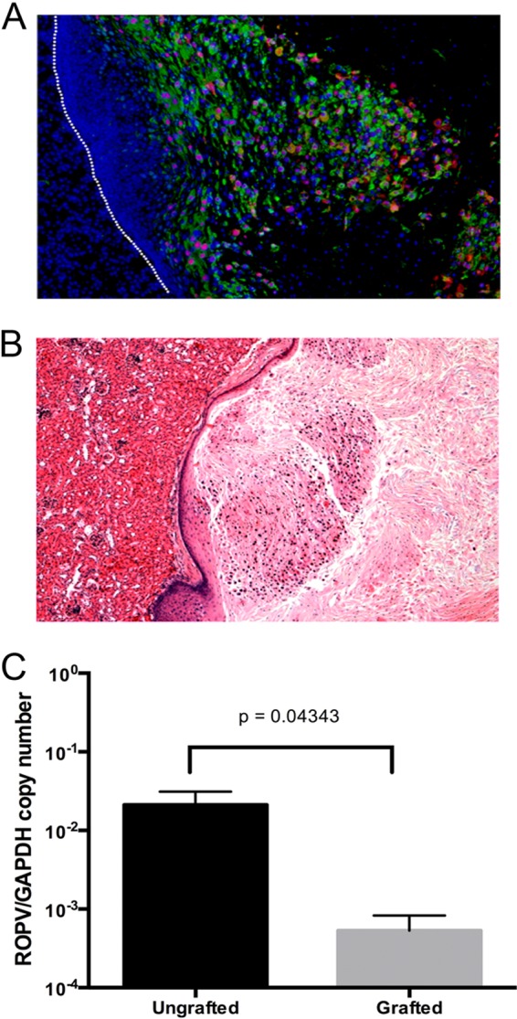
(A) Successful completion of the ROPV life cycle in infected tongue epithelium grafted under the kidney capsules of SCID mice was confirmed by immunostaining for the viral E4 protein (green) and by fluorescence in situ hybridization to detect ROPV DNA (red). (B) Analysis of productively infected tissue from SCID mice revealed a characteristic papilloma pathology in grafted epithelium. The images in panels A and B were captured with a 20× objective. (C) The characteristic pathology shown in panel B was absent from latently infected tissue, even at 16 weeks postgrafting. The ROPV copy number per copy of GAPDH was higher before grafting (black column, ungrafted), with the copy number declining by 16 weeks under the kidney capsule (gray column, grafted), which is indicative of latent persistence but not reactivation.
References
-
- Jamieson DJ, Duerr A, Burk R, Klein RS, Paramsothy P, Schuman P, Cu-Uvin S, Shah K. 2002. Characterization of genital human papillomavirus infection in women who have or who are at risk of having HIV infection. Am. J. Obstet. Gynecol. 186:21–27 - PubMed
-
- Moscicki AB, Ellenberg JH, Vermund SH, Holland CA, Darragh T, Crowley-Nowick PA, Levin L, Wilson CM. 2000. Prevalence of and risks for cervical human papillomavirus infection and squamous intraepithelial lesions in adolescent girls: impact of infection with human immunodeficiency virus. Arch. Pediatr. Adolesc. Med. 154:127–134 - PubMed
-
- Ozsaran AA, Ates T, Dikmen Y, Zeytinoglu A, Terek C, Erhan Y, Ozacar T, Bilgic A. 1999. Evaluation of the risk of cervical intraepithelial neoplasia and human papilloma virus infection in renal transplant patients receiving immunosuppressive therapy. Eur. J. Gynaecol. Oncol. 20:127–130 - PubMed
-
- Palefsky JM, Minkoff H, Kalish LA, Levine A, Sacks HS, Garcia P, Young M, Melnick S, Miotti P, Burk R. 1999. Cervicovaginal human papillomavirus infection in human immunodeficiency virus-1 (HIV)-positive and high-risk HIV-negative women. J. Natl. Cancer Inst. 91:226–236 - PubMed
-
- Paternoster DM, Cester M, Resente C, Pascoli I, Nanhorngue K, Marchini F, Boccagni P, Cillo U, Ribaldone R, Amoruso E, Cocca N, Cuccolo V, Bertolino M, Surico N, Stratta P. 2008. Human papilloma virus infection and cervical intraepithelial neoplasia in transplanted patients. Transplant. Proc. 40:1877–1880 - PubMed
Publication types
MeSH terms
Grants and funding
LinkOut - more resources
Full Text Sources
Other Literature Sources
