. 2014 Feb;88(3):1433-46.
doi: 10.1128/JVI.01815-13. Epub 2013 Oct 30.

Apolipoprotein E codetermines tissue tropism of hepatitis C virus and is crucial for viral cell-to-cell transmission by contributing to a postenvelopment step of assembly

Affiliations

Apolipoprotein E codetermines tissue tropism of hepatitis C virus and is crucial for viral cell-to-cell transmission by contributing to a postenvelopment step of assembly

Kathrin Hueging et al. J Virol. 2014 Feb.

Abstract

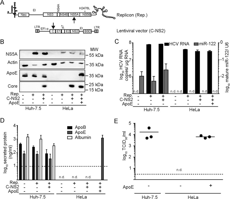
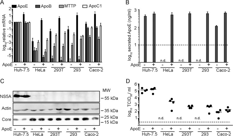
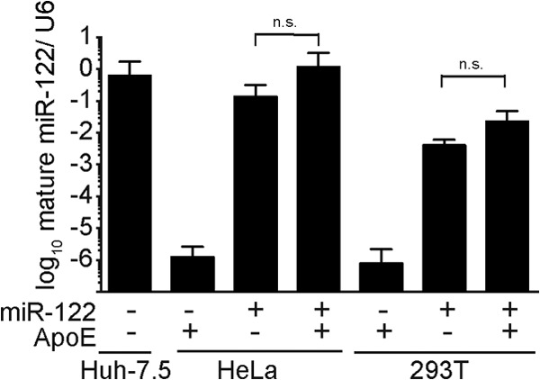
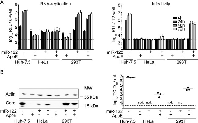
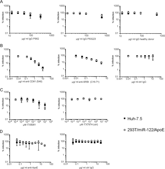
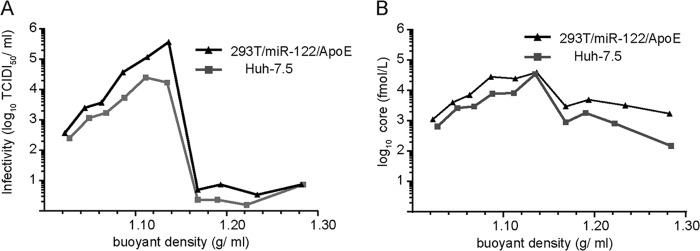
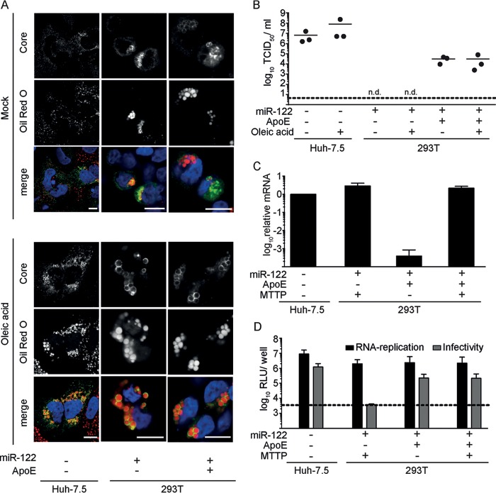
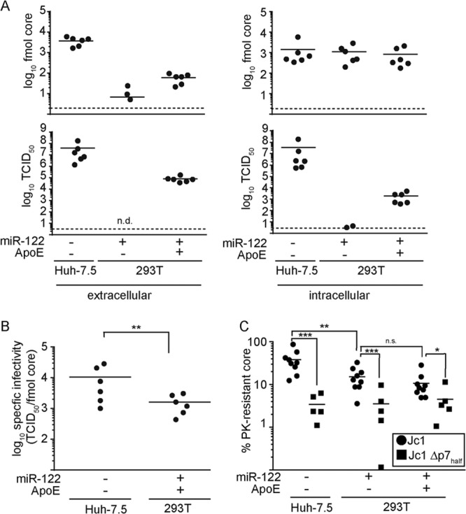
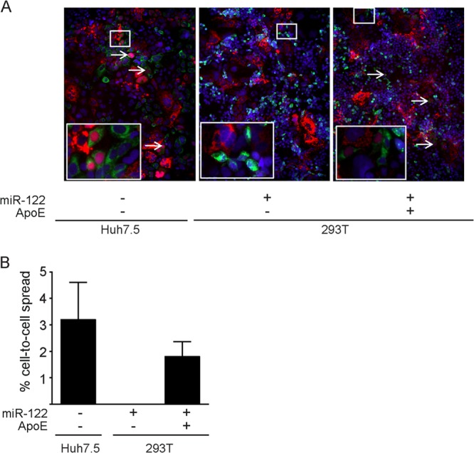
Hepatitis C virus (HCV) predominantly infects human hepatocytes, although extrahepatic virus reservoirs are being discussed. Infection of cells is initiated via cell-free and direct cell-to-cell transmission routes. Cell type-specific determinants of HCV entry and RNA replication have been reported. Moreover, several host factors required for synthesis and secretion of lipoproteins from liver cells, in part expressed in tissue-specific fashion, have been implicated in HCV assembly. However, the minimal cell type-specific requirements for HCV assembly have remained elusive. Here we report that production of HCV trans-complemented particles (HCVTCP) from nonliver cells depends on ectopic expression of apolipoprotein E (ApoE). For efficient virus production by full-length HCV genomes, microRNA 122 (miR-122)-mediated enhancement of RNA replication is additionally required. Typical properties of cell culture-grown HCV (HCVcc) particles from ApoE-expressing nonliver cells are comparable to those of virions derived from human hepatoma cells, although specific infectivity of virions is modestly reduced. Thus, apolipoprotein B (ApoB), microsomal triglyceride transfer protein (MTTP), and apolipoprotein C1 (ApoC1), previously implicated in HCV assembly, are dispensable for production of infectious HCV. In the absence of ApoE, release of core protein from infected cells is reduced, and production of extracellular as well as intracellular infectivity is ablated. Since envelopment of capsids was not impaired, we conclude that ApoE acts after capsid envelopment but prior to secretion of infectious HCV. Remarkably, the lack of ApoE also abrogated direct HCV cell-to-cell transmission. These findings highlight ApoE as a host factor codetermining HCV tissue tropism due to its involvement in a late assembly step and viral cell-to-cell transmission.


Figures

FIG 1
FIG 1
Ectopic expression of Apo E rescues production of infectious HCV in HeLa cells. (A) Schematic depiction of the constructs used for trans-complementation. The replicon (Rep.) harboring a neomycin resistance gene and the adaptive mutation H2476L is shown at the top, and the lentiviral vector (C-NS2) is shown at the bottom. The arrows indicate which gene products were detected in panel B to confirm expression of both constructs in Huh-7.5 and HeLa cells. (B) Protein expression of the given cell lysates was determined using Western blotting and antibodies against NS5A, core, and actin. +, ectopic expression of the constructs; −, only endogenous levels are present. (C) Cells were seeded in 6-well dishes, and 48 h later, total RNA was extracted to determine the amount of HCV RNA and mature miR-122 via RT-PCR. Shown are values normalized to amounts of GAPDH mRNA or U6 RNA, respectively. n.d., not detected. (D) Amounts of secreted ApoE, ApoB, and albumin were determined by collecting culture supernatants at 48 h postseeding and using specific ELISAs. The dashed line shows the detection limit of the assays. (E) HCVTCP titers in the supernatants of the different cell lines were determined at 48 h postseeding using a limiting-dilution assay.
FIG 2
FIG 2
Ectopic expression of ApoE is necessary and sufficient to permit HCV assembly in several human nonliver cell lines. Huh-7.5, HeLa, 293T, 293, and Caco-2 cells were engineered to express HCV C-NS2 proteins and ApoE or the empty vector as a control. Subsequently, these cells were transiently transfected with a subgenomic HCV replicon. Analyses were carried out at 48 h postelectroporation. +, ectopic expression of the different constructs; −, only endogenous levels are present. The dashed lines in panels B and D show the detection limit. n.d., not detected. (A) Total cellular RNA was extracted, and mRNA levels of ApoE, ApoC1, MTTP, and ApoB were determined via mRNA-specific RT-PCR and normalized to GAPDH mRNA. Values were normalized to the corresponding mRNA abundance in Huh-7.5 C-NS2 cells. (B) Secretion of ApoE into the cell culture fluid of the given cell lines was quantified by specific ELISA. (C) Core, NS5A, and actin were detected in cell lysates by Western blotting. (D) Release of infectious HCVTCP from the different cell lines was quantified by a limiting-dilution assay.
FIG 3
FIG 3
Abundance of mature miR-122 in different human cell lines. Levels of mature miR-122 in Huh-7.5, HeLa, and 293T cells were determined via specific RT-PCR and are shown relative to U6 RNA. +, indicates ectopic expression of miR-122; −, only endogenous levels are present; n.s., nonsignificant.
FIG 4
FIG 4
Expression of ApoE and miR-122 is sufficient for production of full-length HCV particles from 293T and HeLa cells. +, ectopic expression of the indicated constructs; −, only endogenous levels are present. The dashed lines in panel A and the right panel of panel B show the detection limit of the assay. n.d., not detected. (A) The indicated cell lines were transfected with JcR2A HCV RNA, which encodes the Renilla luciferase reporter. HCV RNA replication was determined at 4 h, 24 h, 48 h, and 72 h postelectroporation by luciferase measurement and is expressed as relative light units (left). Additionally, cell-free culture fluids of the different cell lines were used to inoculate Huh-7.5 cells, and infectivity was determined by luciferase assay at 48 h postinoculation (right). (B) The same set of cells was transfected with wild-type Jc1 RNA, and at 48 h postelectroporation, the abundance of core protein in the cell lysates was determined by Western blotting (left). Actin served as a loading control. Release of infectious particles was determined by using a limiting-dilution assay (right).
FIG 5
FIG 5
Susceptibility of 293T-derived and Huh-7.5-derived HCV particles to neutralizing antibodies and entry inhibitors. Reporter viruses encoding a Renilla luciferase were produced in Huh-7.5 and in 293T/miR-122/ApoE cells. Cell culture supernatant was harvested at 48 h and 72 h posttransfection. For neutralization and inhibitor treatments, the virus stocks were normalized for Renilla counts prior to infection. Normalized values are shown. (A) Neutralization with patient-derived IgGs. Patient-derived IgGs were purified from HCV-positive donors. HCV-negative donors served as control. (B) Neutralization with anti-CD81 and anti-SCARB1 antibodies and control IgG. (C) Susceptibility to the SCARB1 inhibitor ITX5061; a nonfunctional compound (ITX7874) served as a control. (D) Neutralization with anti-ApoE antibodies.
FIG 6
FIG 6
Buoyant densities of HCV particles derived from 293T/miR-122/ApoE and Huh-7.5 cells. Equilibrium density centrifugation was performed on concentrated cell culture supernatants. The density was determined by refractometry and is given on the x axis. A representative gradient is shown. (A) The infectivity in the different fractions of the gradient was quantified by a limiting-dilution assay. (B) Core amounts were determined by core-specific ELISA.
FIG 7
FIG 7
Lipid droplets and MTTP are not limiting for HCV assembly in 293T/miR-122/ApoE cells. +, ectopic expression; −, only endogenous levels are present. The dashed lines in panels B and D represent the detection limit of the assay. n.d., not detected. (A) The indicated cell lines were transfected with Jc1 RNA, and at 30 h postelectroporation, cells were stimulated with oleic acid (final concentration, 360 μM) or vehicle control overnight. Lipid droplets were stained with Oil Red O (red), core protein was detected by specific antibodies (green), and cell nuclei were visualized with DAPI (blue). Representative pictures are shown, and the scale bar corresponds to 10 μm. (B) The virus titer in the cell culture fluid was determined by using a limiting dilution assay. (C) The indicated cell lines were transfected with JcR2a HCV RNA. Total RNA was extracted at 48 h posttransfection, and mRNA levels of MTTP were determined in the different cell lines by RT-PCR. (D) HCV RNA replication in these cells was determined at 48 h posttransfection by luciferase measurement. Additionally, cell-free culture fluids were used to inoculate naive Huh-7.5 cells. Infectivity was determined by luciferase assay at 48 h postinoculation. Luciferase values are expressed as relative light units (RLU) per 6-well plate (for RNA replication) or per 12-well plate (for infectivity).
FIG 8
FIG 8
ApoE is required prior to particle release and after capsid envelopment. +, ectopic expression of the constructs; −, only endogenous levels are present. (A) At 48 h after transfection of Jc1 RNA into the different cell lines, viral titers and core protein were determined in the cell culture supernatants (left) as well as in freeze-thaw lysates (right). Shown are the total amount of core protein (upper panel)s and the viral titer (lower panels) per 10-cm culture dish. n.d., not detected. The dashed line represents the detection limit of the assay. (B) The specific infectivities of particles in the supernatant, from Huh-7.5 and 293T/miR-122/ApoE cells were calculated based on the data presented in panel A. (C) Cells were lysed by freezing and thawing at 48 h postelectroporation with Jc1 or Jc1 Δp7half RNA and subjected to proteinase K digestion. Lysates were divided into three parts: the first part was left untreated, the second was incubated with proteinase K, and the third was lysed with Triton prior to proteinase K-mediated digestion. The amounts of core protein in the different samples were determined by core-specific ELISA. Normalized values for proteinase K-treated samples are shown. For this, the core amounts in the samples treated with proteinase K and Triton were set as background and those in the untreated samples were set as 100%.
FIG 9
FIG 9
ApoE is required for HCV cell-to-cell transmission. 293T cells expressing miR-122 and ApoE or the empty vector and Huh-7.5 cells were transfected with Jc1 RNA. After electroporation, the cells were cocultured with Huh-7.5 cells harboring an RFP reporter construct that translocates into the nucleus upon cleavage by the HCV protease NS3-4A. Cell-free infection was blocked by agarose overlay and by addition of 20 mg/ml AR4A antibody. (A) After 72 h, cells were fixed with PFA, stained for NS5A, and analyzed by fluorescence microscopy. Representative images are shown. The inset is an enlarged version of a part of the picture. White arrows indicate cells where the RFP reporter translocated to the nucleus. (B) Cell-to-cell transmission of HCV was quantified by counting the total number of cells and the percentage of cells in which the RFP reporter translocated to the nucleus. Shown are averages from 3 experiments. In each experiment, between 1,000 and 2,000 cells were analyzed.

Similar articles

Cited by

References

    1. Lavanchy D. 2011. Evolving epidemiology of hepatitis C virus. Clin. Microbiol. Infect. 17:107–115. 10.1111/j.1469-0691.2010.03432.x - DOI - PubMed
    1. Delang L, Neyts J, Vliegen I, Abrignani S, Neddermann P, De Francesco R. 2013. Hepatitis C virus-specific directly acting antiviral drugs. Curr. Top. Microbiol. Immunol. 369:289–320. 10.1007/978-3-642-27340-7_12 - DOI - PubMed
    1. Seeff LB. 2002. Natural history of chronic hepatitis C. Hepatology 36:S35–S46. 10.1002/hep.1840360706 - DOI - PubMed
    1. Yamane D, McGivern DR, Masaki T, Lemon SM. 2013. Liver injury and disease pathogenesis in chronic hepatitis C. Curr. Top. Microbiol. Immunol. 369:263–288. 10.1007/978-3-642-27340-7_11 - DOI - PubMed
    1. Weissenborn K, Tryc AB, Heeren M, Worthmann H, Pflugrad H, Berding G, Bokemeyer M, Tillmann HL, Goldbecker A. 2009. Hepatitis C virus infection and the brain. Metab. Brain Dis. 24:197–210. 10.1007/s11011-008-9130-5 - DOI - PubMed

Publication types

LinkOut - more resources