. 2014 Jan;25(1):87-94.
doi: 10.1091/mbc.E13-06-0326. Epub 2013 Oct 30.

Substrate stiffness regulates solubility of cellular vimentin

Affiliations

Substrate stiffness regulates solubility of cellular vimentin

Maria E Murray et al. Mol Biol Cell. 2014 Jan.

Abstract

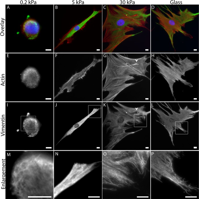
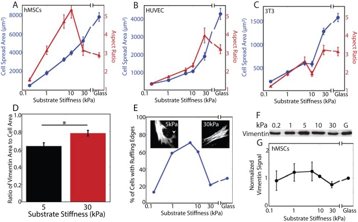
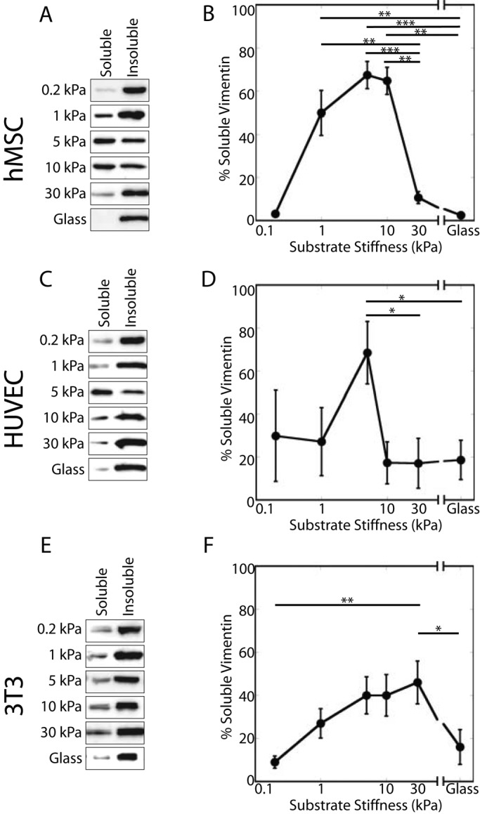
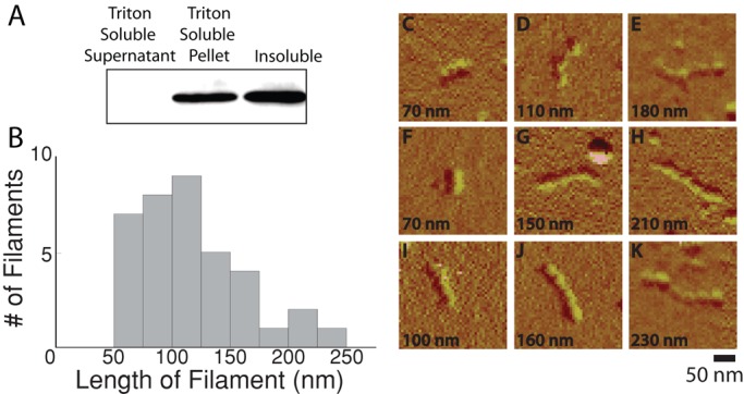
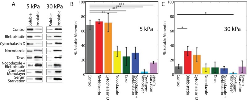
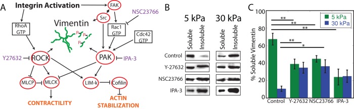
The intermediate filament protein vimentin is involved in the regulation of cell behavior, morphology, and mechanical properties. Previous studies using cells cultured on glass or plastic substrates showed that vimentin is largely insoluble. Although substrate stiffness was shown to alter many aspects of cell behavior, changes in vimentin organization were not reported. Our results show for the first time that mesenchymal stem cells (hMSCs), endothelial cells, and fibroblasts cultured on different-stiffness substrates exhibit biphasic changes in vimentin detergent solubility, which increases from nearly 0 to 67% in hMSCs coincident with increases in cell spreading and membrane ruffling. When imaged, the detergent-soluble vimentin appears to consist of small fragments the length of one or several unit-length filaments. Vimentin detergent solubility decreases when these cells are subjected to serum starvation, allowed to form cell-cell contacts, after microtubule disruption, or inhibition of Rac1, Rho-activated kinase, or p21-activated kinase. Inhibiting myosin or actin assembly increases vimentin solubility on rigid substrates. These data suggest that in the mechanical environment in vivo, vimentin is more dynamic than previously reported and its assembly state is sensitive to stimuli that alter cellular tension and morphology.


Figures

FIGURE 1:
FIGURE 1:
Vimentin in hMSCs on different-stiffness gels. Vimentin networks are evident in cells on gels over a wide range of stiffnesses (A–D; actin, red; vimentin, green; and nuclei, blue). On 0.2-kPa gels hMSCs are small and round (Figure 2, A, E, I, and M). Cells elongate on 5-kPa gels (B, F, J, N). On 30-kPa gels and silanized glass, cells spread similarly and contain stress fibers (G, H), and the vimentin network extends to the cell edge (O, P). Bars, 10 μm.
FIGURE 2:
FIGURE 2:
Substrate stiffness causes changes in spread area, aspect ratio, and ruffling behavior but not total vimentin protein by whole-cell lysis. In all cell types tested, spread area increases with substrate stiffness (A–C; blue), whereas the aspect ratio peaks and then decreases in hMSCs (A; red) and HUVECs (B; red) but not 3T3 cells (C; red). The vimentin network extends to fill a greater proportion of the cell on stiffer substrates (D; 5 kPa, 63 ± 10% vs. 30 kPa, 78 ± 3%; p < 0.05; see also Figure 4, A–D). Changes in ruffling behavior also accompany substrate stiffness changes (E; arrowhead in left inset indicates a ruffling edge; compare to nonruffling cell, right inset; F-actin staining). Total vimentin expression is not altered by substrate stiffness (F, G; p > 0.1 across all conditions).
FIGURE 3:
FIGURE 3:
Detergent-soluble vimentin varies with substrate stiffness. The soluble and insoluble fractions of vimentin in hMSCs, HUVECs, and NIH-3T3 cells vary with substrate stiffness. In all cell types tested, vimentin solubility exhibits a biphasic response characterized by a small soluble pool on the softest substrates, which increases with substrate stiffness until peaking at 5 kPa (A, B, hMSCs; C, D, HUVECs) or 30 kPa (E, F; NIH-3T3 cells).
FIGURE 4:
FIGURE 4:
Detergent solubility and vimentin assembly state. To understand the differences between detergent-soluble vimentin and soluble vimentin as determined by centrifugation, the detergent-soluble vimentin pool was centrifuged at 3000 × g for 10 min. Detergent-soluble vimentin pellets at low speeds when subjected to low-speed centrifugation (A). Quantification showed (n = 3) that 65% of the vimentin was present in the low-detergent pellet, with 3% of the vimentin in the low-detergent supernatant and the majority of the protein in the soluble fraction. The length of the filaments in the detergent-soluble pool was determined by imaging the filaments with AFM (B). Filaments demonstrate a beaded morphology and a diameter similar to previously observed results for vimentin (C–K).
FIGURE 5:
FIGURE 5:
Changes in cytoskeletal organization affect vimentin detergent solubility in hMSCs. Perturbing microtubules decreases the amount of detergent-soluble vimentin in cells on 5-kPa but not 30-kPa gels (A, B; nocodazole, Taxol). In contrast, inhibiting myosin activity or actin assembly increases vimentin detergent solubility in cells on 30-kPa gels (A, C; blebbistatin, cytochalasin D). Inhibiting myosin activity in the presence of depolymerized MT does not compound the effect of MT depolymerization alone (compare nocodazole + blebbistatin to nocodazole).
FIGURE 6:
FIGURE 6:
The effect of signaling inhibition on vimentin detergent solubility is substrate dependent. Both RhoA and Rac1 signaling are important in both mechanotransduction signaling and vimentin phosphorylation (A). ROCK (Y-27632) or Rac1 (NSC23766) inhibition decreases the proportion of detergent-soluble vimentin in cells on 5-kPa gels but increases it in cells on 30-kPa gels (B, C). Inhibiting PAK (IPA-3) decreases vimentin detergent solubility on 5-kPa gels but not on 30-kPa gels (B,C, comparison of control to IPA-3, p = 0.088).

References

    1. Ando S, Nakao K, Gohara R, Takasaki Y, Suehiro K, Oishi Y. Morphological analysis of glutaraldehyde-fixed vimentin intermediate filaments and assembly-intermediates by atomic force microscopy. Biochim Biophys Acta. 2004;1702:53–65. - PubMed
    1. Bhadriraju K, Yang M, Ruiz SA, Pirone DM, Tan J, Chen CS, Alom Ruiz S. Activation of ROCK by RhoA is regulated by cell adhesion, shape, and cytoskeletal tension. Exp Cell Res. 2007;313:3616–3623. - PMC - PubMed
    1. Blikstad I, Lazarides E. Vimentin filaments are assembled from a soluble precursor in avian erythroid cells. J Cell Biol. 1983;96:1803–1808. - PMC - PubMed
    1. Bocquet A, Berges R, Frank R, Robert P, Peterson AC, Eyer J. Neurofilaments bind tubulin and modulate its polymerization. J Neurosci. 2009;29:11043–11054. - PMC - PubMed
    1. Byfield FJ, Reen RK, Shentu T-P, Levitan I, Gooch KJ. Endothelial actin and cell stiffness is modulated by substrate stiffness in 2D and 3D. J Biomech. 2009;42:1114–1119. - PMC - PubMed

Publication types