. 2013 Oct 30;280(1773):20132242.
doi: 10.1098/rspb.2013.2242. Print 2013 Dec 22.

Bacterial farming by the fungus Morchella crassipes

Affiliations

Bacterial farming by the fungus Morchella crassipes

Martin Pion et al. Proc Biol Sci. .

Abstract

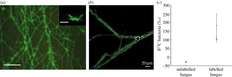
The interactions between bacteria and fungi, the main actors of the soil microbiome, remain poorly studied. Here, we show that the saprotrophic and ectomycorrhizal soil fungus Morchella crassipes acts as a bacterial farmer of Pseudomonas putida, which serves as a model soil bacterium. Farming by M. crassipes consists of bacterial dispersal, bacterial rearing with fungal exudates, as well as harvesting and translocation of bacterial carbon. The different phases were confirmed experimentally using cell counting and (13)C probing. Common criteria met by other non-human farming systems are also valid for M. crassipes farming, including habitual planting, cultivation and harvesting. Specific traits include delocalization of food production and consumption and separation of roles in the colony (source versus sink areas), which are also found in human agriculture. Our study evidences a hitherto unknown mutualistic association in which bacteria gain through dispersal and rearing, while the fungus gains through the harvesting of an additional carbon source and increased stress resistance of the mycelium. This type of interaction between fungi and bacteria may play a key role in soils.

Keywords: Morchella crassipes; Pseudomonas putida; dispersal; exudate consumption; mutualism; sclerotia and melanization.


Figures

Figure 1.
Figure 1.
Benefits of the bacterial partner. (a) Dispersal of P. putida on the mycelium of M. crassipes. The insert shows dispersion of bacteria alone. Scale bars represent 500 µm. (b) Confocal microscopy showing maximum intensity projections of migrating GFP-labelled P. putida cells (fluorescence) on the fungal hyphae of M. crassipes (reflection). (c) Bacterial biomarkers (cy17 : 0, filled circles and 16 : 1ω7, open circles) were significantly 13C-enriched during the rearing phase compared to a control with an unlabelled fungus.
Figure 2.
Figure 2.
Influence of P. putida on the formation of sclerotia. (a) In the absence of bacteria, sclerotia are formed homogeneously throughout the entire surface of the Petri dish. (b) When the fungus is co-inoculated with bacteria, sclerotia are formed only in the region opposed to the bacterial inoculum zone. (c) Sclerotia are homogeneously distributed if the bacterial inoculation area is replaced by depleted agar medium. (d,e) Sclerotia (arrows) are always formed at the most distant position from the bacterial inoculum regardless of its position (stars).
Figure 3.
Figure 3.
Bacterial populations during dispersal and the formation of sclerotia. (a) Bacterial population increase and decrease are linked first to the migration on the fungal mycelium and later to the formation of sclerotia by the fungus. Bacterial populations were counted in three zones of the Petri dish (zones indicated in (c)). Bacteria colonized the entire Petri dish after 5 days, reaching a density close to 7×10⁷ CFUs. However, bacteria in the inoculum zone disappeared during sclerotial formation. (b) In the absence of sclerotia formation, bacteria are dispersed as in previous experiments, but they are not harvested as shown by the absence of a decrease in bacterial population in the inoculum zone. (c) Comparison of fungal phenotype in the presence of bacteria but in the absence of sclerotia formation (ii). The letters indicate the regions for which bacterial populations were counted. (d) In technical agar, bacterial population in the inoculum zone is significantly lower than in purified agar (unpaired t-test on log-transformed data, n = 8, t = 12.16, p < 10−8).
Figure 4.
Figure 4.
Benefits of the fungal partner. (a) The fungal biomarker (18 : 2ω6) was significantly 13C-enriched during the harvesting phase with respect to the same experiment carried out with unlabelled bacteria. (b) Representative images (three replicates per condition) of M. crassipes co-inoculated or not with P. putida and showing the difference in melanization of the fungal mycelium after three weeks of growth. In the presence of P. putida (1, 2 and 3), the mycelium is less dark than in its absence (4, 5 and 6). This difference in colour is attributed to the level of hyphae melanization. 1 and 4 = one-week-old medium; 2 and 5 = 23 min drying; 3 and 6 = 15 min drying.

References

    1. O'Donnell AG, Young IM, Rushton SP, Shirley MD, Crawford JW. 2007. Visualization, modelling and prediction in soil microbiology. Nat. Rev. Microbiol. 5, 689–699 (doi:10.1038/nrmicro1714) - DOI - PubMed
    1. Torsvik V, Ovreas L, Thingstad TF. 2002. Prokaryotic diversity: magnitude, dynamics, and controlling factors. Science 296, 1064–1066 (doi:10.1126/science.1071698) - DOI - PubMed
    1. Gans J, Wolinsky M, Dunbar J. 2005. Computational improvements reveal great bacterial diversity and high metal toxicity in soil. Science 309, 1387–1390 (doi:10.1126/science.1112665) - DOI - PubMed
    1. de Boer W, Folman LB, Summerbell RC, Boddy L. 2005. Living in a fungal world: impact of fungi on soil bacterial niche development. FEMS Microbiol. Rev. 29, 795–811 (doi:10.1016/j.femsre.2004.11.005) - DOI - PubMed
    1. Nazir R, Warmink JA, Boersma H, van Elsas JD. 2010. Mechanisms that promote bacterial fitness in fungal-affected soil microhabitats. FEMS Microbiol. Ecol. 71, 169–185 (doi:10.1111/j.1574-6941.2009.00807.x) - DOI - PubMed