. 2013 Oct 30;5(209):209ra152.
doi: 10.1126/scitranslmed.3006839.

Spherical nucleic acid nanoparticle conjugates as an RNAi-based therapy for glioblastoma

Affiliations

Spherical nucleic acid nanoparticle conjugates as an RNAi-based therapy for glioblastoma

Samuel A Jensen et al. Sci Transl Med. .

Abstract

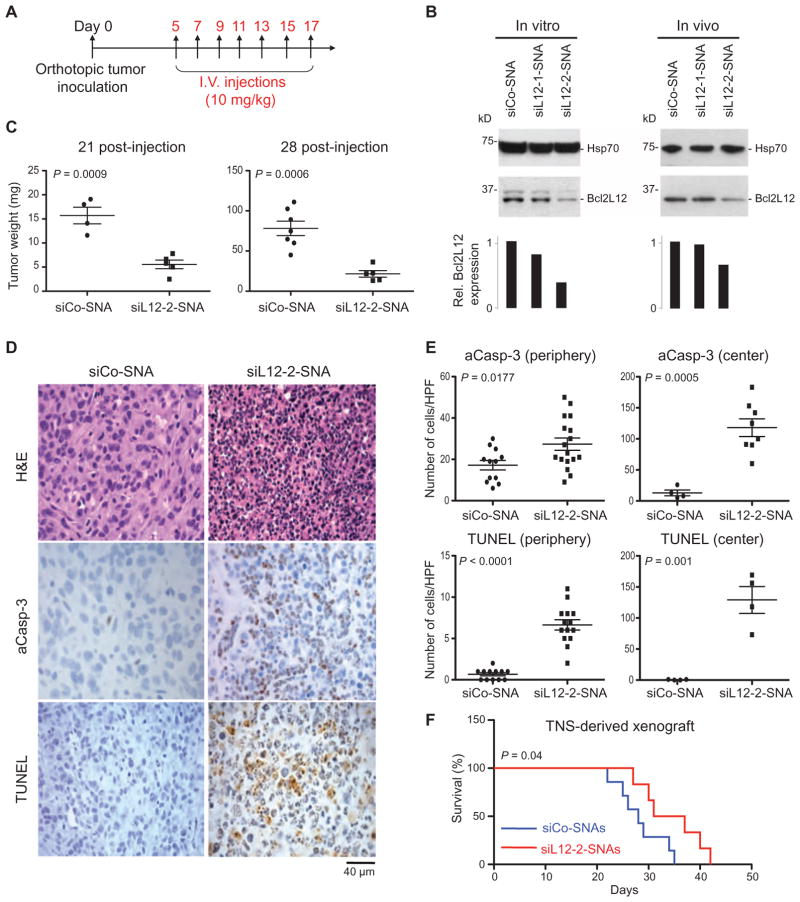
Glioblastoma multiforme (GBM) is a neurologically debilitating disease that culminates in death 14 to 16 months after diagnosis. An incomplete understanding of how cataloged genetic aberrations promote therapy resistance, combined with ineffective drug delivery to the central nervous system, has rendered GBM incurable. Functional genomics efforts have implicated several oncogenes in GBM pathogenesis but have rarely led to the implementation of targeted therapies. This is partly because many "undruggable" oncogenes cannot be targeted by small molecules or antibodies. We preclinically evaluate an RNA interference (RNAi)-based nanomedicine platform, based on spherical nucleic acid (SNA) nanoparticle conjugates, to neutralize oncogene expression in GBM. SNAs consist of gold nanoparticles covalently functionalized with densely packed, highly oriented small interfering RNA duplexes. In the absence of auxiliary transfection strategies or chemical modifications, SNAs efficiently entered primary and transformed glial cells in vitro. In vivo, the SNAs penetrated the blood-brain barrier and blood-tumor barrier to disseminate throughout xenogeneic glioma explants. SNAs targeting the oncoprotein Bcl2Like12 (Bcl2L12)--an effector caspase and p53 inhibitor overexpressed in GBM relative to normal brain and low-grade astrocytomas--were effective in knocking down endogenous Bcl2L12 mRNA and protein levels, and sensitized glioma cells toward therapy-induced apoptosis by enhancing effector caspase and p53 activity. Further, systemically delivered SNAs reduced Bcl2L12 expression in intracerebral GBM, increased intratumoral apoptosis, and reduced tumor burden and progression in xenografted mice, without adverse side effects. Thus, silencing antiapoptotic signaling using SNAs represents a new approach for systemic RNAi therapy for GBM and possibly other lethal malignancies.


Conflict of interest statement

Competing interests: D.A.G., C.A.M., W.L.D., A.H.S., and P.C.P. have interest in AuraSense Therapeutics, which develops SNA-based technologies. The content is solely the responsibility of the authors and does not necessarily represent the official views of the sponsors or government, and no official endorsement should be inferred.

Figures

Fig. 1
Fig. 1. SNAs penetrate glial cells and down-regulate Bcl2L12 in vitro
(A) Uptake of Cy5-labeled SNAs (red) into huTNS and the human glioma cell line U87MG. Cells were colabeled with fluorescein isothiocyanate (FITC)–conjugated cytochalasin (green) and 4′,6-diamidino-2-phenylindole (DAPI) (blue) to visualize actin filaments and nuclei, respectively. Scale bars, 40 μm. BF, bright field. (B) Effect of Bcl2L12-targeting SNAs siL12-1 and siL12-2 on Bcl2L12 protein levels in huTNS, LN235, and LNZ308 glioma cells relative to control (siCo-SNA) cultures as assessed by Western blot using the noted concentrations of SNAs. Hsp70 served as a loading control. Bar graphs represent a densitometric analysis of Western blots. Band intensity was normalized to Hsp70 and then expressed relative to Bcl2L12 levels in siCo-treated samples. (C) Bcl2L12 knockdown persistance in LN235 cells treated with 1 nM siCo-SNA, siL12-1-SNA, or siL12-2-SNA for 48, 72, 96, and 120 hours as assessed by Western blot. (D) Schematic depicting siL12-2 siRNA cleavage site and subsequent 5′-RLM-RACE PCR. (E) Resulting bands from successive rounds of PCR on the cDNA template using GeneRacer- and Bcl2L12-specific primers in siL12-2-SNA–and siCo-SNA–treated cells. The arrow points to the cleavage product. The asterisk represents a PCR product that is a fragment of Bcl2L12, not ligated to the GeneRacer oligo, and is thus a nonspecific band. (F) DNA sequence chromatogram of resulting PCR product from siL12-2-SNA–treated cells.
Fig. 2
Fig. 2. Bcl2L12-specific SNAs promote apoptotic signaling in glioma cells
(A and B) Bcl2L12 knockdown by siL12-SNAs sensitizes cells to apoptosis (measured by caspase-3 and caspase-7 cleavage) upon treatment with staurosporine (STS) and temozolomide (TMZ). LS, large subunit; LS+N, large subunit plus N-peptide. (C) Effect of Bcl2L12 knockdown on p53 protein stability, phosphorylated p53 (p-p53Ser15), and the p53 target p21 upon doxorubicin (Doxo) treatment for the indicated times. (D) p53 target gene induction of p21 on the mRNA level as measured by quantitative reverse transcription polymerase chain reaction (qRT-PCR). (E) Bcl2L12 mRNA knockdown was assessed in parallel and expressed as fold change relative to siCo-SNA–treated cells at 0, 4, 8, and 16 hours after application of doxorubicin. Data are means ± SD (n = 3).
Fig. 3
Fig. 3. SNAs disseminate throughout glioma tissue
(A) ICP-MS quantification of SNA uptake into orthotopic U87MG tumor and adjacent normal tissue after 48 hours. Data are means ± SEM (n = 3 glioma-bearing mice, n = 5 normal mice). P value was calculated with one-way analysis of variance (ANOVA). (B) Schematic overview of the synthesis of Gd(III)-functionalized SNAs. (i) Gd(III)-SNA conjugates were prepared from alkyne-modified T bases and azide-labeled Gd(III) complexes through click chemistry. (ii) Gd(III)-conjugated DNA was then functionalized onto gold nanoparticle (Au-NP) surface to form Gd(III)-SNA following the same procedure as fig. S3, except without oligoethylene glycol/polyethylene glycol (PEG) backfill. (C) MR images of tumor-bearing mouse brains injected intracranially with Gd(III)-SNAs. Two representative coronal sections imaged 24 hours after Gd(III)-SNA injection (upper panel) show localization of Gd(III)-SNA within the intracerebral lesion. Gd(III) signal is white and outlined in black dotted line. Also shown are corresponding hematoxylin and eosin (H&E) sections showing the location of the tumor (darker purple, outlined in light gray dotted line), and three-dimensional (3D) reconstruction of MR images [Gd(III) signal in red]. See movie S1. (D) LA-ICP-MS shows localization of Au, Fe, and Gd(III) contents in coronal brain sections of mice injected intracranially with Gd(III)-SNAs. The heat map indicates the relative amount of the element detected in the tissue. (E) Confocal fluorescence microscopy of coronal brain sections derived from tumor-bearing and non–tumor-bearing mice injected with saline or Cy5-SNAs. Representative sections are shown for n = 5 mice.
Fig. 4
Fig. 4. SNAs cross the BBB/BTB and selectively accumulate in glioma tissue
(A) Noncontact in vitro BBB model using a coculture of huBMECs and human astrocytes. Representative confocal fluorescence microscopy images demonstrate Cy5.5-SNA (red) distribution in endothelial and astrocytic cells. Endothelial and astrocytic cells stained positively for occludin (a marker for tight junctions) and glial fibrillary acidic protein (GFAP), respectively. Anti-vimentin and DAPI stained cytoplasm and nuclei, respectively. (B and C) IVIS analysis of brains with or without U87MG (B) or huTNS (C) tumors 48 hours after systemic delivery of saline or Cy5.5-SNAs. SNA accumulation is indicated by increased fluorescence (yellow). Quantification of radiant efficiency is shown as relative signal amount under the images. (D and E) Selective accumulation of Cy5.5-SNAs within U87MG (D) or huTNS (E) tumors, but not normal brain elements. Coronal brain sections were silver-stained and counterstained with hematoxylin. Yellow arrows indicate sites of SNA accumulation, which appear as dark brown regions. T, tumor; N, normal brain. (F) huTNS-derived accumulated SNAs in vascular and extravascular tumor elements as revealed by silver staining and anti-CD31 immunohistochemistry on adjacent coronal sections. Arrows point to areas of SNA and CD31 colocalization. Hashed box shows SNAs that extravasated into the tumor parenchyma.
Fig. 5
Fig. 5. Systemic administration of SNAs reduces intratumoral Bcl2L12 and decreases tumor burden in mice
(A) Scheme of cell and SNA injections. Mice were orthotopically injected with either U87MG or huTNS cells for tumor burden, histology, and survival analyses (day 0) followed by seven intravenous (I.V.) injections of siCo- or siL12-SNAs. (B) Bcl2L12 knockdown in U87MG-derived intracranial tumors treated with siL12-SNAs. Hsp70 was used as a loading control. Histograms represent a densitometric analysis of the Western blots. The intensity of Bcl2L12 bands was normalized to Hsp70 and ex-pressedasrelativeBcl2L12 expression. (C) Weight of xenografted tumors 21 and 28 days after SNA injection. Data points display tumor weight from individual mice. P value was calculated with two-tailed Student’s t test. (D and E) Intratumoral apoptosis in mice injected with siL12-2-SNA. (E) The amount of activated caspase-3 (aCasp-3) and terminal deoxynucleotidyl transferase–mediated deoxyuridine triphosphate nick end labeling (TUNEL) was quantified from (D) in peripheral and central tumor regions. Data points are the number of stained cells per field. Bars are means ± SEM. P values were calculated with two-tailed Student’s t test. HPF, high-power field. (F) Kaplan-Meier survival curves of mice with TNS-derived xenografts treated with siL12-2-SNA (n = 6) or siCo-SNA (n = 7). P value was calculated with the Mantel-Cox test.

Comment in

References

    1. Dunn GP, Rinne ML, Wykosky J, Genovese G, Quayle SN, Dunn IF, Agarwalla PK, Chheda MG, Campos B, Wang A, Brennan C, Ligon KL, Furnari F, Cavenee WK, Depinho RA, Chin L, Hahn WC. Emerging insights into the molecular and cellular basis of glioblastoma. Genes Dev. 2012;26:756–784. - PMC - PubMed
    1. Furnari FB, Fenton T, Bachoo RM, Mukasa A, Stommel JM, Stegh A, Hahn WC, Ligon KL, Louis DN, Brennan C, Chin L, DePinho RA, Cavenee WK. Malignant astrocytic glioma: Genetics, biology, and paths to treatment. Genes Dev. 2007;21:2683–2710. - PubMed
    1. Wen PY, Kesari S. Malignant gliomas in adults. N Engl J Med. 2008;359:492–507. - PubMed
    1. Engelman JA, Settleman J. Acquired resistance to tyrosine kinase inhibitors during cancer therapy. Curr Opin Genet Dev. 2008;18:73–79. - PubMed
    1. Stukel JM, Caplan MR. Targeted drug delivery for treatment and imaging of glioblastoma multiforme. Expert Opin Drug Deliv. 2009;6:705–718. - PubMed

Publication types

MeSH terms