. 2013 Dec 13;288(50):35769-80.
doi: 10.1074/jbc.M113.511212. Epub 2013 Oct 30.

Suppression of lysosome function induces autophagy via a feedback down-regulation of MTOR complex 1 (MTORC1) activity

Affiliations

Suppression of lysosome function induces autophagy via a feedback down-regulation of MTOR complex 1 (MTORC1) activity

Min Li et al. J Biol Chem. .

Abstract

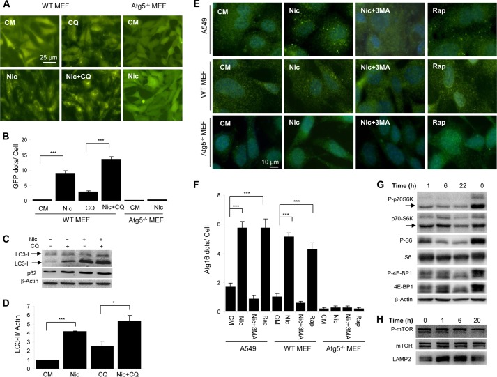
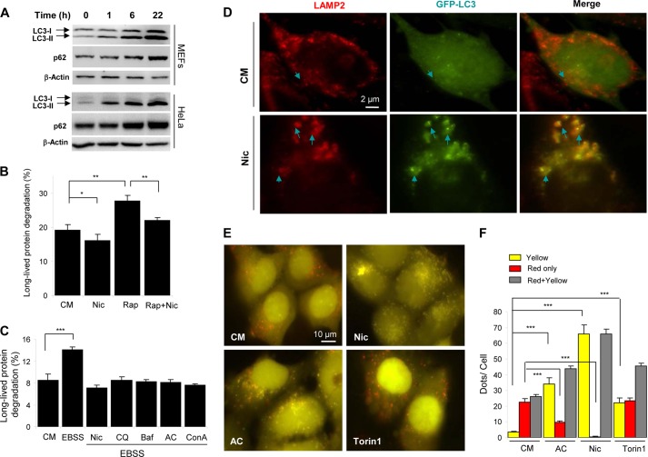
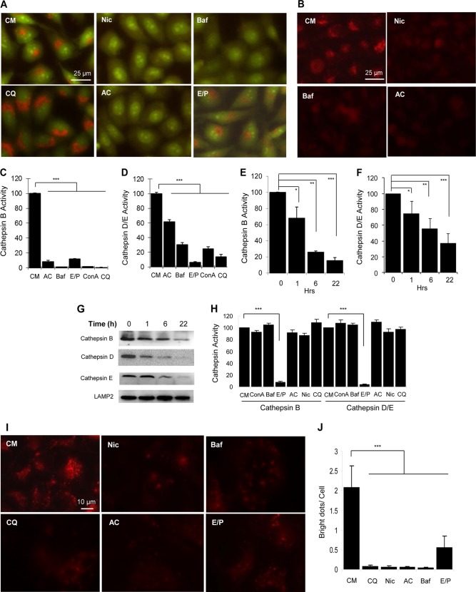
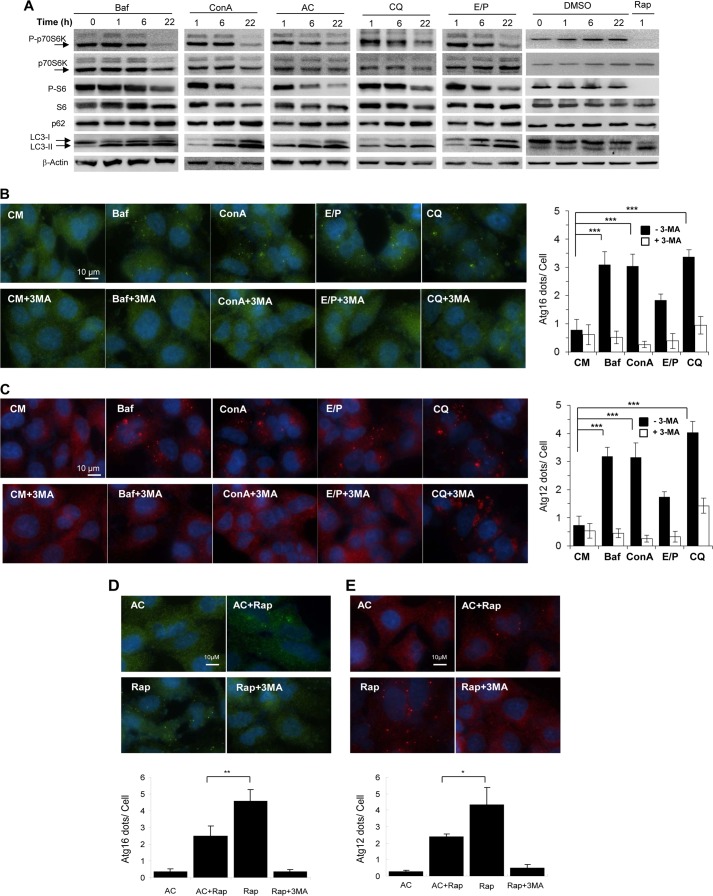
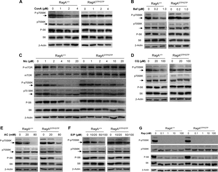
Autophagy can be activated via MTORC1 down-regulation by amino acid deprivation and by certain chemicals such as rapamycin, torin, and niclosamide. Lysosome is the degrading machine for autophagy but has also been linked to MTORC1 activation through the Rag/RRAG GTPase pathway. This association raises the question of whether lysosome can be involved in the initiation of autophagy. Toward this end, we found that niclosamide, an MTORC1 inhibitor, was able to inhibit lysosome degradation and increase lysosomal permeability. Niclosamide was ineffective in inhibiting MTORC1 in cells expressing constitutively activated Rag proteins, suggesting that its inhibitory effects were targeted to the Rag-MTORC1 signaling system. This places niclosamide in the same category of bafilomycin A1 and concanamycin A, inhibitors of the vacuolar H(+)-ATPase, for its dependence on Rag GTPase in suppression of MTORC1. Surprisingly, classical lysosome inhibitors such as chloroquine, E64D, and pepstatin A were also able to inhibit MTORC1 in a Rag-dependent manner. These lysosome inhibitors were able to activate early autophagy events represented by ATG16L1 and ATG12 puncta formation. Our work established a link between the functional status of the lysosome in general to the Rag-MTORC1 signaling axis and autophagy activation. Thus, the lysosome is not only required for autophagic degradation but also affects autophagy activation. Lysosome inhibitors can have a dual effect in suppressing autophagy degradation and in initiating autophagy.

Keywords: Autophagy; Cell Biology; Lysosomes; Signal Transduction; mTOR.


Figures

FIGURE 1.
FIGURE 1.
Niclosamide induces autophagy and inhibition of MTORC1. A and B, wild type and Atg5-deficient MEFs expressing GFP-LC3 were treated with or without niclosamide (Nic; 10 μm) and CQ (40 μm) for 6 h and then assessed for GFP-LC3 puncta formation (A), which was quantified (B). C and D, as treated in A, MEFs were then lysed for immunoblot analysis (C). Densitometry was performed, and the ratio of LC3-II/β-actin was determined (D). E and F, wild type and Atg5-deficient MEFs or A549 cells were treated as indicated (Nic, 10 μm; 3-MA, 10 mm; Rap, 1 μm) for 6 h, followed by immunostaining using an anti-ATG16L1 antibody (E). ATG16L1 puncta were quantified (F). G and H, HeLa cells were treated with niclosamide (10 μm) for different times as indicated. The zero time point represents cells untreated and harvested at the end of the incubation period. Total lysates (G) or the lysosome-enriched heavy membrane fraction (H) was subjected to immunoblot analysis. For B, D, and F, values represent means ± S.D. from three independent experiments. ***, p < 0.001; *, p < 0.05. CM, complete medium.
FIGURE 2.
FIGURE 2.
Niclosamide inhibits autophagic degradation. A, MEFs or HeLa cells were treated with niclosamide (Nic; 10 μm) for 0–22 h as indicated, followed by immunoblot analysis for p62 and LC3. B and C, MEFs were incubated in complete medium (CM), or in Earle's balanced salt solution (EBSS) with or without niclosamide (10 μm), CQ (40 μm), Baf (0.5 μm), AC (40 mm) or ConA (2.5 μm) for 6 h (C), or incubated in complete medium (CM) with or without niclosamide (10 μm) and rapamycin (Rap; 1 μm) for 16 h (B). The long-lived protein degradation was measured. D, GFP-LC3-expressing MEFs were treated as indicated (Nic, 10 μm) for 6 h, followed by immunostaining with anti-LAMP2. Arrows indicated colocalized GFP-LC3 (green) and LAMP2 (red) puncta. E and F, HEK-293A cells expressing GFP-RFP-LC3 were treated as indicated (Nic, 10 μm; AC, 40 mm; or Torin1, 250 nm) for 6 h (E). Puncta showing both green and red fluorescence (indicated as yellow) or showing only the red fluorescence (indicated as red) were quantified (F). Values represent means ± S.D. from three independent experiments. ***, p < 0.001; **, p < 0.01; *, p < 0.05.
FIGURE 3.
FIGURE 3.
Niclosamide inhibits lysosomal degradation capacity. A and B, HeLa cells were treated with niclosamide (Nic; 10 μm), CQ (40 μm), Baf (0.5 μm), AC (40 mm), or E64D (25 μm) plus pepstatin A (50 μm) (E/P) for 6 h, followed by staining with AO (1 μg/ml, A) or LTR (50 ng/ml, B) for 30 min. C–G, HeLa cells were treated with various chemicals (40 mm AC; 0.5 μm Baf; 25 μm/50 μm E64D/pepstatin A; 2.5 μm ConA; 40 μm CQ) as indicated (C and D) for 6 h, or with niclosamide (10 μm) for 0 to 20 h (E–G). The lysosome-enriched cytosolic fraction was analyzed for cathepsin B (C and E) and cathepsin D/E (D and F) activities and for immunoblot analysis (G). Cathepsin activities were standardized to that of the untreated sample, which was set at the 100% level. H, whole cell lysates of untreated HeLa cells (4 μg) were mixed with the various chemicals at the concentrations indicated above for 1 h. The activities of cathepsin B and cathepsin D/E were measured. I and J, HeLa cells were preincubated with DQ-BSA(10 μg/ml) for 1 h and then treated as in A (I). The degraded products presented as red puncta, which were quantified (J). For C–F, H, and J, values represent means ± S.D. from three independent experiments. ***, p < 0.001; **, p < 0.01; *, p < 0.05. CM, complete medium.
FIGURE 4.
FIGURE 4.
Lysosome inhibitors can inhibit MTORC1 and initiate autophagosome formation. A, MEFs were treated with Baf (0.5 μm), ConA (2.5 μm), AC (40 mm), CQ (40 μm), or E64D (25 μm) plus pepstatin A (50 μm) (E/P) for 0 to 22 h, followed by immunoblot analysis. B and C, A549 cells were treated as indicated with or without 3-MA (10 mm) for 6 h, followed by immunostaining with anti-ATG16L1 (B) or anti-ATG12 (C). D and E, A549 cells were treated as indicated (40 mm AC; 1 μm rapamycin; 10 mm 3-MA) for 6 h, followed by immunostaining with anti-ATG16L1 (D) or anti-ATG12 (E), which were quantified. For panels D and E, values represent means ± S.D. from three independent experiments. **, p < 0.01; *, p < 0.05. DMSO, dimethyl sulfoxide; CM, complete medium; Rap, rapamycin.
FIGURE 5.
FIGURE 5.
Inhibition of MTORC1 activity by lysosome inhibitors is regulated by Rag GTPases. Wild type (RagA+/+) and RagAGTP/GTP knock-in MEF cells were cultured in Earle's balanced salt solution with ConA (A), Baf (B), niclosamide (Nic; C), CQ (D), AC (E), E64D plus pepstatin A (E/P) (F) or rapamycin (G) at different concentrations as indicated for 1 h. Cells were then cultured in complete medium (CM) containing the same level of chemicals for an additional hour. Cell lysates were prepared and subjected to immunoblot assays with the indicated antibodies.

References

    1. Yang Z., Klionsky D. J. (2010) Eaten alive: a history of macroautophagy. Nat. Cell Biol. 12, 814–822 - PMC - PubMed
    1. Jung C. H., Ro S. H., Cao J., Otto N. M., Kim D. H. (2010) mTOR regulation of autophagy. FEBS Lett. 584, 1287–1295 - PMC - PubMed
    1. Fleming A., Noda T., Yoshimori T., Rubinsztein D. C. (2011) Chemical modulators of autophagy as biological probes and potential therapeutics. Nat. Chem. Biol. 7, 9–17 - PubMed
    1. Yip C. K., Murata K., Walz T., Sabatini D. M., Kang S. A. (2010) Structure of the human mTOR complex I and its implications for rapamycin inhibition. Mol. Cell 38, 768–774 - PMC - PubMed
    1. Balgi A. D., Fonseca B. D., Donohue E., Tsang T. C., Lajoie P., Proud C. G., Nabi I. R., Roberge M. (2009) Screen for chemical modulators of autophagy reveals novel therapeutic inhibitors of mTORC1 signaling. PLoS One 4, e7124. - PMC - PubMed

Publication types

MeSH terms

Substances