. 2013 Dec 13;288(50):35913-24.
doi: 10.1074/jbc.M113.529727. Epub 2013 Oct 30.

Identification of a novel role of ZMIZ2 protein in regulating the activity of the Wnt/β-catenin signaling pathway

Affiliations

Identification of a novel role of ZMIZ2 protein in regulating the activity of the Wnt/β-catenin signaling pathway

Suk Hyung Lee et al. J Biol Chem. .

Abstract

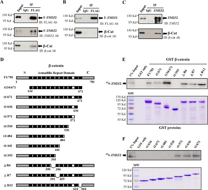
ZMIZ2, also named ZIMP7, is a protein inhibitor of activated STAT (PIAS)-like protein and a transcriptional coactivator. In this study, we investigated the interaction between ZMIZ2 and β-catenin, a key regulator of the Wnt signaling pathway. We demonstrated that the expression of exogenous ZMIZ2 augments TCF (T cell factor) and β-catenin-mediated transcription. In contrast, shRNA knockdown of ZMIZ2 expression specifically represses the enhancement of TCF/β-catenin-mediated transcription by ZMIZ2. Using Wnt3a-conditioned medium, we demonstrated that ZMIZ2 can enhance Wnt ligand-induced TCF/β-catenin-mediated transcription. We also showed a promotional role of ZMIZ2 in enhancing β-catenin downstream target gene expression in human cells and in Zmiz2 null (Zmiz2(-/-)) mouse embryonic fibroblasts (MEFs). The regulatory role of Zmiz2 in Wnt-induced TCF/β-catenin-mediated transcription can be restored in Zmiz2(-/-) MEFs that were infected with adenoviral expression vectors for Zmiz2. Moreover, enhancement of Zmiz2 on TCF/β-catenin-mediated transcription was further demonstrated in Zmiz2 knockout and Axin2 reporter compound mice. Furthermore, the protein-protein interaction between ZMIZ2 and β-catenin was identified by co-immunoprecipitation and in vitro protein pulldown assays. We also observed recruitment of endogenous ZMIZ2 onto the promoter region of the Axin 2 gene, a β-catenin downstream target promoter, in a Wnt ligand-inducible manner. Finally, a promotional role of ZMIZ2 on cell growth was demonstrated in human cell lines and Zmiz2 knockout MEFs. Our findings demonstrate a novel interaction between ZMIZ2 and β-catenin and elucidate a novel mechanism for PIAS-like proteins in regulating Wnt signaling pathways.

Keywords: Cell Signaling; PIAS-like Proteins; Protein-Protein Interactions; Transcription Regulation; Wnt Ligand; Wnt Signaling; ZMIZ2; β-Catenin.


Figures

FIGURE 1.
FIGURE 1.
ZMIZ2 controls β-catenin/TCF-mediated transcription. A, 100 ng of pGL3-OT (OT-Luc) or pGL3-OF (OF-Luc), 25 ng of pcDNA3-β-gal, 5 ng of TCF1 expression vector, 5 ng of β-catenin (β-cat), 10 ng of pcDNA3-FLAG-ZMIZ1, and 10 ng of pcDNA3-FLAG-ZMIZ2 were transfected into HEK293 cells. Cells were cultured for 24 h, and luciferase and β-gal activities were measured. B, 100 ng of pGL3-OT or pGL3-OF, 25 ng of pcDNA3-β-gal and/or 5 ng of TCF1 expression vector, 5 ng of β-catenin, 10 ng of pcDNA3-FLAG-ZMIZ2, and other shRNA constructs, as indicated in the figures, were transfected into HEK293 cells. Luciferase and β-gal activities were measured. Similar experiments were repeated with the TCF4 expression vector in HEK293 cells C, luciferase activity is reported as relative light units (luciferase/β-galactosidase) and represented as the mean ± S.D. *, p < 0.05; **, p < 0.01 (see text).
FIGURE 2.
FIGURE 2.
ZMIZ2 regulates cyclin D1 promoter activity and Wnt-induced β-catenin/TCF-mediated transcription. A, 100 ng of pGL3-Cyclin D1 promoter (Cyclin D1-Luc), 25 ng of pcDNA3-β-gal, and various amount of pcDNA3-FLAG-ZMIZ2, pcDNA3-FLAG-ZMIZ1, and other shRNA constructs, as indicated, were transfected into HEK293 cells. Cells were cultured for 24 h in the regular media, and luciferase and β-gal activities were measured. Similar experiments were repeated with LNCaP cells (B). C, 100 ng of pGL3-OT (OT-Luc) or pGL3-OF (OF-Luc), 25 ng of pcDNA3-β-gal, 5 ng of TCF4 expression vector, 5 ng of β-catenin, and 10 ng of pcDNA3-FLAG-ZMIZ2 were transfected into HEK293 cells. Cells were cultured for 24 h, washed, and incubated with either Wnt3a-CM or L-CM for another 24 h. The cells were harvested, and luciferase and β-gal activities were measured. Luciferase activity is reported as relative light units (luciferase/β-galactosidase) and represented as the mean ± S.D. *, p < 0.05; **, p < 0.01 (see text).
FIGURE 3.
FIGURE 3.
ZMIZ2 is required for the expression of β-catenin target genes in response to Wnt3a. HEK293 cells were infected with a lentivirus encoding shRNA against ZMIZ2 or GFP. The infected cells were cultured for 72 h and then harvested for Western blot assays with either anti-ZMIZ2 or anti-tubulin antibody (A) or incubated for a further 6 h in the presence of Wnt3a-CM or L-CM and then harvested for quantitative RT-PCR (B). The levels of cyclin D1, c-Myc, and Axin2 were normalized to that of GAPDH mRNA. C, MEFs were prepared from different genotype embryos at E10.5. Whole cell lysates were analyzed by Western blot assays with either anti-ZMIZ2 or anti-tubulin antibody. D, MEFS were cultured in the presence of Wnt3a-CM or L-CM for 6 h and then harvested for quantitative RT-PCR. The levels of Axin2, c-Myc, c-Jun, and Cd44 were normalized to that of GAPDH mRNA. E and F, different genotypes of MEFs were infected with an adenovirus expressing GFP (Adv GFP) or FLAG-ZMIZ2 (Adv Zmiz2). The infected cells were cultured for 24 h, harvested for Western blot assays with anti-ZMIZ2, anti-FLAG, anti-GFP, or anti-tubulin antibody (E), or incubated for a further 6 h in the presence of Wnt3a-CM or L-CM and then harvested for quantitative RT-PCR (F). The levels of Axin2, c-Myc, and cyclin D1 were measured and normalized to that of GAPDH mRNA. The relative mRNA levels from each sample are presented as the mean ± S.D. of triplicate reactions. *, p < 0.05 (see text).
FIGURE 4.
FIGURE 4.
Loss of ZMIZ2 inhibits cell growth. A, HEK293 cells were infected with a lentivirus encoding shRNA against ZMIZ2 or GFP. The infected cells were cultured for 4 and 6 days and then analyzed by Western blot assays with either anti-ZMIZ2 or anti-tubulin antibody. Infected HEK293 cells were seeded into 96-well plates and cultured in the regular media (B) or in the presence of Wnt3a-CM or L-CM (C). Then, cell growth was measured every other day by MTS assay. ZMIZ2 shRNA or GFP shRNA encoding lentivirus-infected LAPC4 cells (D) and LNCaP cells (F) were cultured, and then cell growth was measured by MTS assay. E, for the colony formation assay, ∼500 LAPC4 cells infected with either GFP shRNA or ZMIZ2 shRNA were seeded in 6-well plates. After 12 days, cells were fixed and stained with crystal violet. G, MTS cell assay using E10.5 MEFs isolated from Zmiz2 heterozygous intercrosses (+/+, n = 3; +/−, n = 4; −/−, n = 3). The data represent the mean ± S.D. of three independent experiments. *, p < 0.05 (see text).
FIGURE 5.
FIGURE 5.
Physical interaction between ZMIZ2 and β-catenin in vitro and in intact cells. A, HEK293 cells were transfected with pcDNA3-FLAG-ZMIZ2 and pcDNA3-β-catenin. Cell lysates were then immunoprecipitated (IP) with the anti-FLAG antibody (Ab) or normal mouse IgG and then analyzed on SDS-PAGE with anti-FLAG, anti-ZMIZ2, or anti-β-catenin (β-cat) antibody. IB, immunoblot. B, HEK293 cells were transfected with pcDNA3-FLAG-ZMIZ1 and pcDNA3-β-catenin. Cell lysates were immunoprecipitated with the anti-FLAG antibody or normal mouse IgG and then analyzed on SDS-PAGE with anti-FLAG or anti-β-catenin antibody. C, whole cell lysates of HEK293 cells were immunoprecipitated with the anti-ZMIZ2 antibody or normal rabbit IgG and analyzed on SDS-PAGE with anti-ZMIZ2 or anti-β-catenin antibody. D, GST fusion proteins containing full length with both the amino terminus (N) and the carboxy terminus (C) regions and different deletion mutants of β-catenin were constructed. E and F, equal amounts of GST fusion proteins containing full-length and different deletion mutants of β-catenin were used to pull down in vitro-translated [35S]methionine-labeled full-length ZMIZ2. GST protein alone was used as a negative control and analyzed with other fusion proteins with molecular weight (MW) markers. Materials bound to glutathione-Sepharose beads were subjected to SDS-PAGE and autoradiography. Equal amounts of GST fusion proteins were resolved in SDS-PAGE and stained with Coomassie Blue for measuring expression.
FIGURE 6.
FIGURE 6.
Alteration of the Axin2 expression in Zmiz2 knockout mice. β-Galactosidase staining of E10.5 embryos using the Wnt activity reporter Axin2LacZ/+. Reduced staining was observed dorsally and in the forebrain, midbrain, hindbrain, and mandibular branches of Axin2LacZ/+:Zmiz2−/− (D) embryos as compared with Axin2LacZ/+:Zmiz2+/+ (C) embryos. No staining was detected in Axin2+/+:Zmiz2−/− (B) and Axin2+/+:Zmiz2+/+ (A) embryos. E, HEK293 cells were cultured in the presence of Wnt3a-CM or L-CM for 1 h. Cells were then cross-linked with formaldehyde, sheared by sonication, and immunoprecipitated with anti-ZMIZ2 antibody, anti-β-catenin antibody, or normal rabbit IgG. Primers specific to the β-catenin/TCF-binding sites (−60, 60 bp before the transcriptional initiation site) in the Axin2 promoter were used to PCR-amplify the eluted chromatin. Primers specific to the GAPDH promoter were used as a control to monitor immunoprecipitation specificity.

References

    1. Shuai K. (2000) Modulation of STAT signaling by STAT-interacting proteins. Oncogene 19, 2638–2644 - PubMed
    1. Megidish T., Xu J. H., Xu C. W. (2002) Activation of p53 by protein inhibitor of activated Stat1 (PIAS1). J. Biol. Chem. 277, 8255–8259 - PubMed
    1. Jackson P. K. (2001) A new RING for SUMO. Wrestling transcriptional responses into nuclear bodies with PIAS family E3 SUMO ligases. Genes Dev. 15, 3053–3058 - PubMed
    1. Kotaja N., Karvonen U., Jänne O. A., Palvimo J. J. (2002) PIAS proteins modulate transcription factors by functioning as SUMO-1 ligases. Mol. Cell Biol. 22, 5222–5234 - PMC - PubMed
    1. Schmidt D., Müller S. (2003) PIAS/SUMO. New partners in transcriptional regulation. Cell Mol. Life Sci. 60, 2561–2574 - PMC - PubMed

Publication types

MeSH terms