. 2014 Jan;111(2):300-12.
doi: 10.1152/jn.00841.2012. Epub 2013 Oct 30.

Activity of long-lead burst neurons in pontine reticular formation during head-unrestrained gaze shifts

Affiliations

Activity of long-lead burst neurons in pontine reticular formation during head-unrestrained gaze shifts

Mark M G Walton et al. J Neurophysiol. 2014 Jan.

Abstract

Primates explore a visual scene through a succession of saccades. Much of what is known about the neural circuitry that generates these movements has come from neurophysiological studies using subjects with their heads restrained. Horizontal saccades and the horizontal components of oblique saccades are associated with high-frequency bursts of spikes in medium-lead burst neurons (MLBs) and long-lead burst neurons (LLBNs) in the paramedian pontine reticular formation. For LLBNs, the high-frequency burst is preceded by a low-frequency prelude that begins 12-150 ms before saccade onset. In terms of the lead time between the onset of prelude activity and saccade onset, the anatomical projections, and the movement field characteristics, LLBNs are a heterogeneous group of neurons. Whether this heterogeneity is endemic of multiple functional subclasses is an open question. One possibility is that some may carry signals related to head movement. We recorded from LLBNs while monkeys performed head-unrestrained gaze shifts, during which the kinematics of the eye and head components were dissociable. Many cells had peak firing rates that never exceeded 200 spikes/s for gaze shifts of any vector. The activity of these low-frequency cells often persisted beyond the end of the gaze shift and was usually related to head-movement kinematics. A subset was tested during head-unrestrained pursuit and showed clear modulation in the absence of saccades. These "low-frequency" cells were intermingled with MLBs and traditional LLBNs and may represent a separate functional class carrying signals related to head movement.

Keywords: PPRF; gaze; head movement; primate; saccade.


Figures

Fig. 1.
Fig. 1.
Schematic representation of behavioral tasks. A: in the delay task, a head-mounted laser and a visual target (T1) were illuminated. The monkey was required to look to and align the laser spot with this target. After a variable fixation period, a 2nd target (T2) was switched on. The animal had to maintain fixation at T1 for the delay period. When T1 and the head-mounted laser were switched off, the animal was permitted to look to the location of T2. Successful fixation of this target resulted in a liquid reward. B: the target blank task was identical to the delay task, except that T2 was extinguished shortly after the initiation of a gaze shift to look to it. The reward was contingent on successful fixation of the spatial location of the (now-extinguished) T2.
Fig. 2.
Fig. 2.
Example trials from a long-lead burst neuron (LLBN) with a high peak firing rate. Three groups of 4 trials are shown, with initial eye positions to the left (left column), centered in the orbits (middle column), or to the right (right column). All columns show rightward gaze shifts with amplitudes between 49° and 51°. The 1st row shows eye (black), gaze (blue), and head (red) position traces. The 2nd row shows velocity traces for each. The 3rd and 4th rows show rasters and cumulative spike histograms for this cell. As the initial eye position becomes more rightward, the head contribution increases, the duration increases, and the peak eye and gaze velocities decrease. Note that the cell consistently ceases discharge shortly before the end of the gaze shift.
Fig. 3.
Fig. 3.
Example trials from a cell with a low peak firing rate. All conventions are the same as Fig. 2. Although the movements are highly similar to the ones shown in Fig 2, this neuron's discharge is quite different. In addition to the much-lower firing rate, note that the cell consistently continues to discharge after the end of the gaze shift.
Fig. 4.
Fig. 4.
Movement field plots for the higher-frequency cell shown in Fig. 2 (A) and the lower-frequency cell shown in Fig. 3 (B). The amplitude of the vertical component of the gaze shift is plotted as a function of the amplitude of the horizontal component. The color of the dots indicates the number of spikes (Num. Sp.). The scale bar applies to A and B. Although the number of spikes is, of course, much higher for the higher-frequency cell, both show a monotonic increase in spike count with horizontal gaze amplitude.
Fig. 5.
Fig. 5.
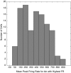
Distribution of peak firing rates (FR). Data for each cell were binned according to horizontal gaze amplitude (bin size = 10°). The mean peak firing rate was computed for each bin. To compare firing rates across cells in a way that would not be affected by differences in the tuning characteristics, the peak firing rate for each cell was taken to be the mean peak firing rate for the bin with the highest mean peak firing rate. The histogram shows the distribution of this value across all of the cells in our sample. Note the large numbers of cells that did not reach 300 spikes/s consistently for gaze shifts of any vector within the range of data collected.
Fig. 6.
Fig. 6.
Median burst lag. A: distribution of median burst lag values for all cells in our sample. B: median burst lag histogram for higher-frequency cells only. Note that an overwhelming majority ceased discharge during the 50-ms period preceding gaze offset. The distribution for lower-frequency cells (C) looks quite different. Most cells in this category continued to discharge after the gaze shift had ended.
Fig. 7.
Fig. 7.
Comparison of burst lag on delay trials and target blank trials. For all cells for which data were available for both delay trials and target blank trials (n = 33), the median burst lag for the target blank trials was plotted against the median burst lag for delay trials. For the target blank trials, a given trial was included in the analysis only if no detectable saccades occurred during the 400-ms period following the end of the primary gaze shift. Open circles indicate lower-frequency cells; filled circles indicate higher-frequency cells. The postgaze activity observed for many of the lower-frequency cells was not dependent on the occurrence of a corrective saccade.
Fig. 8.
Fig. 8.
A: example of a low-frequency neuron showing the relationship between instantaneous firing rate and instantaneous horizontal head velocity. Note that the firing rate never exceeded ∼165 spikes/s. B: comparison of the model fit to the firing rate for 4 individual trials. Black lines = horizontal head velocity; blue lines = firing rate; red lines = predicted firing rate based on a simple model, including only a bias term and horizontal head velocity.
Fig. 9.
Fig. 9.
Example data from a low-frequency cell recorded during both gaze shifts and head-unrestrained pursuit. A: the cell showed a consistent increase in firing rate associated with 50° leftward gaze shifts. It continued to discharge well after the end of the gaze shift until approximately the time the head stopped moving (gray, shaded area). B: four head-unrestrained pursuit trials. The gray, shaded area shows a period of >350 ms, in which the head was moving, but no detectable saccades occurred on any of the trials. The cell consistently discharged during these periods. Note the 1 trial in which the cell began to discharge well in advance of the pursuit movement. This early discharge was observed consistently in association with small, on-direction, “head-only” movements (marked with arrows). C: four-example, head-unrestrained pursuit trials, in which the direction of pursuit reversed unpredictably. Trials are aligned on the point of head-movement reversal. The cell consistently paused during the acceleration phase of the off-direction movement (blue, shaded area) but resumed firing during the deceleration phase.
Fig. 10.
Fig. 10.
Comparison of head-velocity coefficients for dynamic model fits for saccadic gaze shifts and head-unrestrained pursuit. For several neurons, the relationship between firing rate and horizontal head velocity was nearly identical, regardless of whether the head movement accompanied a saccade or pursuit (points along the unity line). For 2 cells, however, the slopes were notably higher for the pursuit condition (∼0.6 vs. ∼0.2).
Fig. 11.
Fig. 11.
Tissue slice showing the site of marking lesions made in monkey P. The dots show reconstructed recording sites for the cells from this monkey. Black dots indicate higher-frequency cells, and red dots indicate lower-frequency cells. Lower- and higher-frequency cells were often recorded in the same track in close proximity to each other and often, in close proximity to medium-lead burst neurons (MLBs). The slice shown was near the caudal edge of the area where MLBs and LLBNs were recorded. To provide a reference point, a 2nd tissue slice is shown (0.5 mm caudal to the 1st slice) in which abducens nucleus (Abd) can be seen.

Similar articles

Cited by

References

    1. Bahill AT, Clark MR, Stark L. The main sequence, a tool for studying human eye movements. Math Biosci 24: 191–204, 1975
    1. Bahill AT, Stark L. Oblique saccadic eye movements. Independence of horizontal and vertical channels. Arch Ophthalmol 95: 1258–1261, 1977 - PubMed
    1. Baloh RW, Konrad HR, Sills AW, Honrubia V. The saccade velocity test. Neurology 25: 1071–1076, 1975a - PubMed
    1. Baloh RW, Sills AW, Kumley WE, Honrubia V. Quantitative measurement of saccade amplitude, duration, and velocity. Neurology 25: 1065–1070, 1975b - PubMed
    1. Becker W. The neurobiology of saccadic eye movements. Metrics Rev Oculomot Res 3: 13–67, 1989 - PubMed

Publication types

LinkOut - more resources