. 2013 Oct 30;33(44):17264-77.
doi: 10.1523/JNEUROSCI.1729-13.2013.

Modulation of NMDAR subunit expression by TRPM2 channels regulates neuronal vulnerability to ischemic cell death

Affiliations

Modulation of NMDAR subunit expression by TRPM2 channels regulates neuronal vulnerability to ischemic cell death

Ishraq Alim et al. J Neurosci. .

Abstract

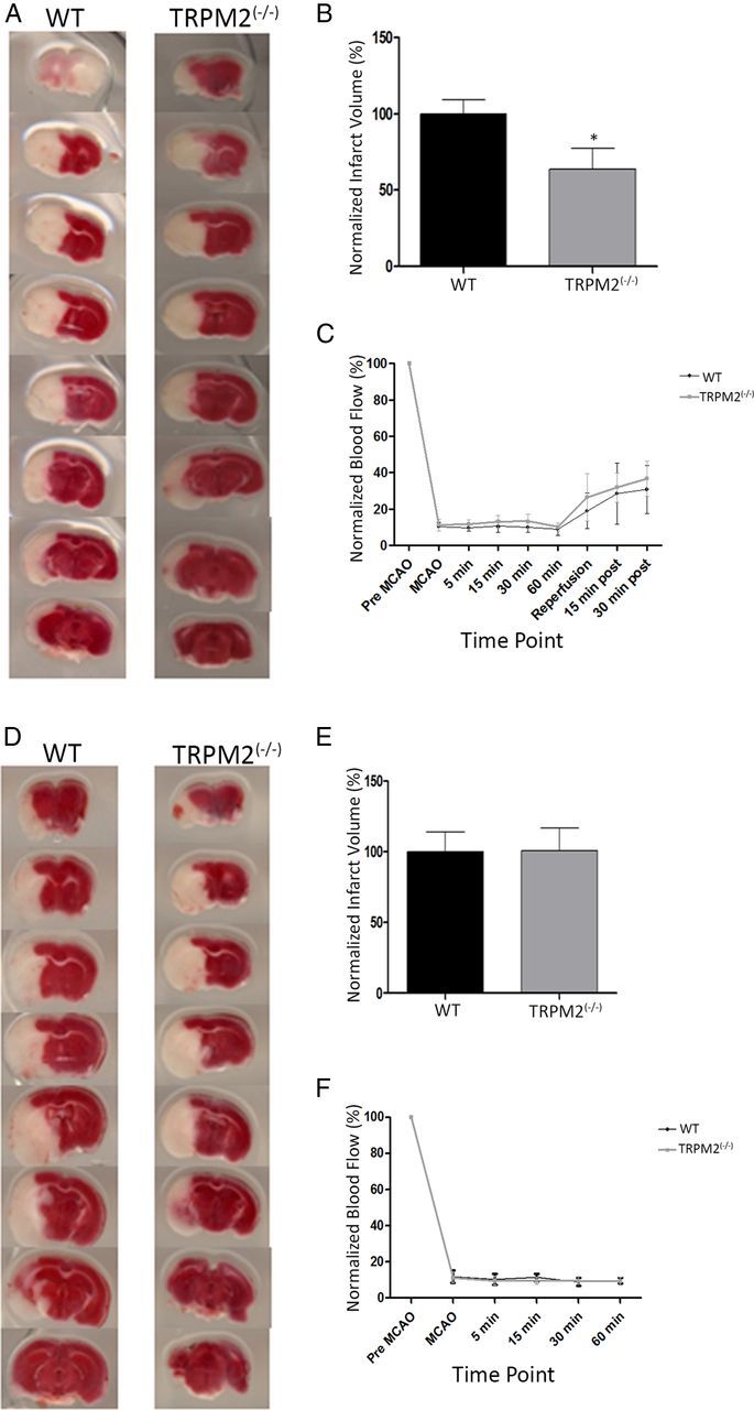
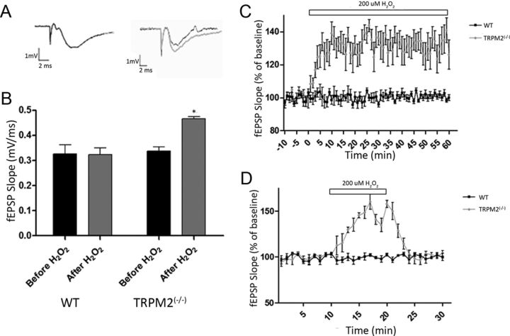
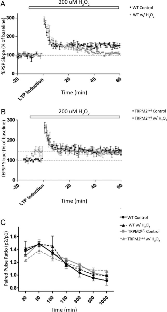
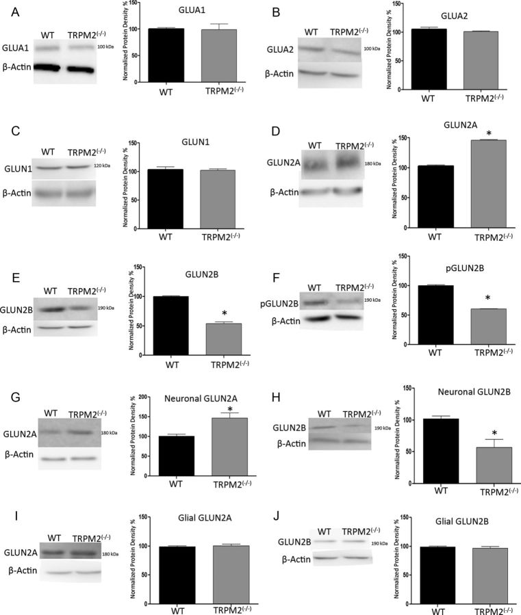
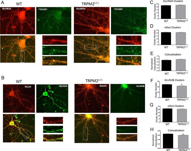
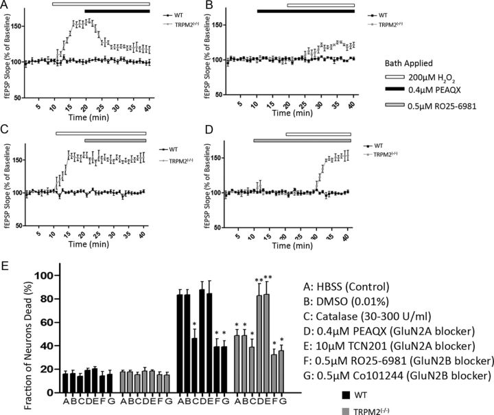
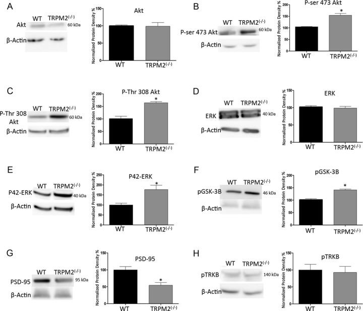
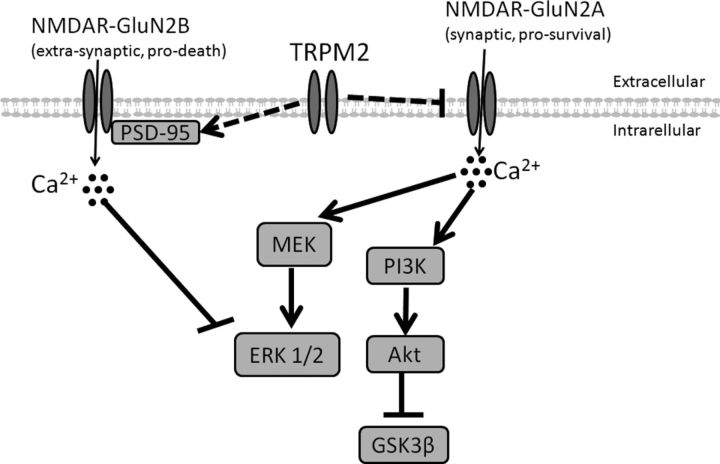
Neuronal vulnerability to ischemia is dependent on the balance between prosurvival and prodeath cellular signaling. In the latter, it is increasingly appreciated that toxic Ca(2+) influx can occur not only via postsynaptic glutamate receptors, but also through other cation conductances. One such conductance, the Transient receptor potential melastatin type-2 (TRPM2) channel, is a nonspecific cation channel having homology to TRPM7, a conductance reported to play a key role in anoxic neuronal death. The role of TRPM2 conductances in ischemic Ca(2+) influx has been difficult to study because of the lack of specific modulators. Here we used TRPM2-null mice (TRPM2(-/-)) to study how TRPM2 may modulate neuronal vulnerability to ischemia. TRPM2(-/-) mice subjected to transient middle cerebral artery occlusion exhibited smaller infarcts when compared with wild-type animals, suggesting that the absence of TRPM2 is neuroprotective. Surprisingly, field potentials (fEPSPs) recorded during redox modulation in brain slices taken from TRPM2(-/-) mice revealed increased excitability, a phenomenon normally associated with ischemic vulnerability, whereas wild-type fEPSPs were unaffected. The upregulation in fEPSP in TRPM2(-/-) neurons was blocked selectively by a GluN2A antagonist. This increase in excitability of TRPM2(-/-) fEPSPs during redox modulation depended on the upregulation and downregulation of GluN2A- and GluN2B-containing NMDARs, respectively, and on augmented prosurvival signaling via Akt and ERK pathways culminating in the inhibition of the proapoptotic factor GSK3β. Our results suggest that TRPM2 plays a role in downregulating prosurvival signals in central neurons and that TRPM2 channels may comprise a therapeutic target for preventing ischemic damage.


Figures

Figure 1.
Figure 1.
TRPM2−/− reduces infarct volume in transient MCAO, but not in permanent MCAO. MCAO was induced in both WT and TRPM2−/− animals. The brains were processed into coronal sections that were stained with TTC. A, B, Infarcts assessed at 48 h after a 1 h tMCAO. Left, Representative coronal sections from WT and TRPM2−/− mice. White areas represent ischemic damage from infarct. Right, Comparison of infarct volumes. There was a reduction of 36.43 ± 16.65% in infarct volumes in the TRPM2−/− animals (n = 8) compared with WT: *p < 0.05. D, E, Infarcts assessed pMCAO. There were no significant differences between infarct volumes in WT and TRPM2−/− animals (n = 5): p > 0.05. C, F, Blood flow was measured using laser Doppler, pMCAO was confirmed for 1 h after occlusion, and reflow was confirmed in tMCAO for up to 30 min after MCAO.
Figure 2.
Figure 2.
H2O2 (200 μm) selectively increases the evoked baseline fEPSP slope in TRPM2−/− neurons. Field potentials (fEPSP) evoked via stimulation in the Schaffer collaterals (at 100–200 μA) were recorded in the hippocampal CA1 region in slices from TRPM2−/− and WT mice under control (ACSF) and oxidative (ACSF+ 200 μm H2O2) conditions. H2O2 was administered through bath application. A, B, Both WT and TRPM2−/− had similar raw fEPSP slope during baseline recordings (black bar); and during H2O2 exposure (gray bar), TRPM2−/− slices showed a 49.0 ± 3.4% increase in fEPSP slope: *p < 0.05. Representative fEPSP recordings (A) using gray and black to represent with and without H2O2, respectively, showed this effect in TRPM2−/−. C, A longer application of H2O2 caused fEPSP to increase in TRPM2−/− (n = 7; gray) for as long as it is applied (60 min), without any effect in WT (n = 5; black). D, The H2O2-induced increase in fEPSP in TRPM2−/− (gray; n = 8) was found to be reversible when application of 200 μm H2O2 (white bar) was removed, with no effect on WT (black; n = 8).
Figure 3.
Figure 3.
H2O2 (200 μm) inhibits LTP formation in both WT and TRPM2−/−. Synaptic plasticity was induced by stimulating the Schaffer collaterals and measuring changes in fEPSPs in the dendrites of CA1 hippocampal neurons. A, B, LTP was induced by HFS, and fEPSPs were recorded both before HFS (for 20 min) and after HFS (for 60 min). H2O2 (200 μm) was applied at 10 min before HFS (white bar). Slices from WT (n = 10; A) and TRPM2−/− (n = 10; B) animals were able to form LTP under control conditions (ACSF; black), and LTP formation was blocked by 200 μm H2O2 (gray) in both WT and TRPM2−/−. Black dotted line indicates baseline fEPSPs under ACSF; gray dotted line indicates baseline of TRPM2−/− with H2O2 (only in B). C, Paired pulse facilitation was induced by giving two pulses with the same amplitude (100–200 μA) at different interpulse intervals. There were no observable changes in paired pulse facilitation between slices from TRPM2−/− and WT animals under both control (ACSF) and with 200 μm H2O2 (n = 10 each group).
Figure 4.
Figure 4.
TRPM2−/− hippocampi have reduced GluN2B expression and increased GluN2A expression. Western blots detecting glutamate receptor expression in hippocampal protein extracts from 3- to 8-week-old TRPM2−/− and WT animals. A, B, Densitometry of protein expression of AMPA subunits GluA1 and GluA2 showed no significant difference between TRPM2−/− and WT (p > 0.05; n = 4 blots with 3 samples of each genotype). C, NMDA subunit GluN1, which binds to GluN2 subunits, also did not have any significant change in protein expression, suggesting that total NMDAR is the same between TRPM2−/− and WT hippocampi. D–F, However, protein expression of NMDAR GluN2 subunits did change, where basal GluN2A expression was increased by 43.30 ± 2.33% in the TRPM2−/− (gray bar) compared with WT (black bar; n = 3; D). *p < 0.01. Both basal and active GluN2B is reduced in TRPM2−/− by 46.21 ± 3.18% and 39.55 ± 1.22%, respectively, compared with WT (n = 3) (E, F). *p < 0.01. G, H, Western blots and densitometry of neuronal cultures from WT and TRPM2−/− mice show that absence of TRPM2 increased GluN2A expression by 47.30 ± 12.57% (n = 3, *p < 0.05) and decreased GluN2B expression by 43.66 ± 12.44% (n = 3, *p < 0.05) compared with WT primary neuronal cultures. I, J, Primary glial cultures showed no change in GluN2A and GluN2B expression (n = 3, p > 0.05).
Figure 5.
Figure 5.
TRPM2−/− reduces synaptic colocalization of GluN2B, but not GluN2A. Immunofluorescence of detecting GluN2A and GluN2B clusters in hippocampal CA1 neurons from WT and TRPM2−/− animals. A, To detect synaptic GluN2A, equal dendritic segments were used to count labeled clusters, where green fluorescence represents synaptic marker VGLUT expression and red represents GluN2A subunit expression. B, Synaptic GluN2B are labeled as follows: red fluorescence is VGLUT, and green is GluN2B. Overlaid images were used to count colocalized clusters. C, WT and TRPM2−/− had no difference in number of GluN2A clusters (n = 27; p > 0.05). D, E, There was also no change in number of synapse and GluN2A synaptic colocalization between neurons from WT and TRPM2−/− animals (n = 27; p > 0.05). F, The number of GluN2B clusters were reduced in hippocampal neurons from TRPM2−/− animals compared with those from WT animals (n = 24). *p < 0.05. G, H, GluN2B synaptic colocalization was also reduced in TRPM2-null neurons compared with WT (n = 24; *p < 0.05), with no effect on total number of synapses. H, Colocalization of GluN2B and VGLUT was reduced in TRPM2−/− compared with WT.
Figure 6.
Figure 6.
GluN2A modulates H2O2-induced excitability in TRPM2−/− hippocampal neurons. fEPSPs evoked by stimulating Schaffer collaterals (100–200 μA) were measured in the CA1 hippocampal dendritic region in slices from TRPM2−/− and WT animals. A, After 10 min of baseline fEPSP recording, 200 μm H2O2 (white bar) was applied to induce an increase in fEPSP slope in TRPM2-null slices, with no effect on WT (as described earlier). Bath application of GluN2A-specific antagonist (0.4 μm of PEAQX, black bar) at 20 min reduced the H2O2-induced increase in fEPSP slope in TRPM2−/− slices (n = 7 p < 0.05), with no effect on WT. B, When GluN2A-specific antagonist was applied at 10 min without H2O2, there was no change in baseline fEPSP slope in both WT and TRPM2−/− slices. At 20 min, 200 μm H2O2 was applied, resulting in a smaller increase (compared with H2O2 only) in fEPSP slope in TRPM2−/− slices and had no effect in WT. C, Application of GluN2B-specific antagonist (0.5 μm RO256981, gray bar) after 10 min of 200 μm H2O2 had no effect on the H2O2-induced increase in fEPSP slope seen in TRPM2−/− slices (n = 7). D, GluN2B- specific antagonist, when applied before H2O2 application, had no effect on baseline fEPSP slope (n = 7) in both TRPM2−/− and WT slices, nor did it have any effect on oxidative stress-induced excitability in TRPM2−/− slices when H2O2 was applied. All compounds were diluted in ACSF. E, After 1 h OGD, neuronal cultures from TRPM2−/− mice had reduced cell death compared with cultures from WT (p < 0.01). The neuroprotection from the lack of TRPM2 was inhibited by application of GluN2A-specific antagonists (PEAQX and TCN201). In WT cultures, catalase, which breaks down H2O2, and GluN2B-specific antagonists (RO25–6981 and Co101244) provided neuroprotection after 1 h OGD. DMSO vehicle had no effect on survival of WT or TRPM2−/− cultures. *Significant difference from WT HBSS (control) 1 h OGD. **Significant difference from TRPM2−/− HBSS (control) 1 h OGD.
Figure 7.
Figure 7.
TRPM2−/− increases activity in ERK and Akt pathways. Western blots of whole hippocampi from WT and TRPM2−/− animals detecting changes in expression of proteins responsible for downstream NMDAR-mediated cell survival mechanisms. A, Basal levels of Akt had no change in expression between WT and TRPM2−/−. B, C, However, phospho-Akt was increased at both phosphorylation sites (49.39 ± 8.26% in Ser-473; 65.11 ± 13.78% in Thr-308; n = 4; *p < 0.01) in TRPM2−/− hippocampi compared with WT. D, There was no change in the basal ERK protein expression comparing WT and TRPM2−/− hippocampi. E, Active ERK was increased by 76.44 ± 23.12% (n = 4; *p < 0.01) in TRPM2−/− compared with WT. F, GSK3β is phosphorylated by Akt; in TRPM2−/− hippocampi, there was a 38.96 ± 4.97% (n = 3; *p < 0.01) increase in phosphorylation of GSK3β compared with WT. G, PSD-95 protein expression was decreased by 45.61 ± 12.76% (n = 3; *p < 0.01) in hippocampus from TRPM2−/− animals compared with WT. H, BDNF-mediated TRKB phosphorylation, which is known to be involved in NMDA-mediated neuroprotection, was not affected by the absence of TRPM2 (n = 3). Together, these results show that the absence of TRPM2 promotes prosurvival pathways in the hippocampus.
Figure 8.
Figure 8.
Proposed mechanism of cell death involving TRPM2. The expression of TRPM2 is required to promote expression of PSD-95 and inhibit GluN2A subunit of NMDA receptors. PSD-95 is responsible for transporting GluN2B-containing NMDAR to the cell surface and activating GluN2B by binding. When PSD95 activates GluN2B-containing NMDAR, there is an influx of extrasynaptic Ca2+, which leads to inhibition of phosphorylation of ERK1/2 and promotes cell death. The inhibition of GluN2A expression reduces synaptic Ca2+ influx and prevents downstream activation of MEK and PI3 kinases, which are required for phosphorylation of ERK1/2 and Akt, respectively. Phosphorylation of Akt inhibits proapoptotic factor GSK3β. Overall, TRPM2 channel inhibits expression of prosurvival NMDARs and increases expression of prodeath NMDARs.

References

    1. Aarts MM, Tymianski M. TRPMs and neuronal cell death. Pflugers Arch. 2005;451:243–249. doi: 10.1007/s00424-005-1439-x. - DOI - PubMed
    1. Aarts M, Liu Y, Liu L, Besshoh S, Arundine M, Gurd JW, Wang YT, Salter MW, Tymianski M. Treatment of ischemic brain damage by perturbing NMDA receptor-PSD-95 protein interactions. Science. 2002;298:846–850. doi: 10.1126/science.1072873. - DOI - PubMed
    1. Aarts M, Iihara K, Wei WL, Xiong ZG, Arundine M, Cerwinski W, MacDonald JF, Tymianski M. A key role for TRPM7 channels in anoxic neuronal death. Cell. 2003;115:863–877. doi: 10.1016/S0092-8674(03)01017-1. - DOI - PubMed
    1. Arundine M, Tymianski M. Molecular mechanisms of glutamate-dependent neurodegeneration in ischemia and traumatic brain injury. Cell Mol Life Sci. 2004;61:657–668. doi: 10.1007/s00018-003-3319-x. - DOI - PMC - PubMed
    1. Auberson YP, Allgeier H, Bischoff S, Lingenhoehl K, Moretti R, Schmutz M. 5-Phosphonomethylquinoxalinediones as competitive NMDA receptor antagonists with a preference for the human 1A/2A, rather than 1A/2B receptor composition. Bioorg Med Chem Lett. 2002;12:1099–1102. doi: 10.1016/S0960-894X(02)00074-4. - DOI - PubMed

Publication types

MeSH terms