. 2013 Oct 30;33(44):17314-25.
doi: 10.1523/JNEUROSCI.2129-13.2013.

Conditional ablation of neuroprogenitor cells in adult mice impedes recovery of poststroke cognitive function and reduces synaptic connectivity in the perforant pathway

Affiliations

Conditional ablation of neuroprogenitor cells in adult mice impedes recovery of poststroke cognitive function and reduces synaptic connectivity in the perforant pathway

Chongran Sun et al. J Neurosci. .

Abstract

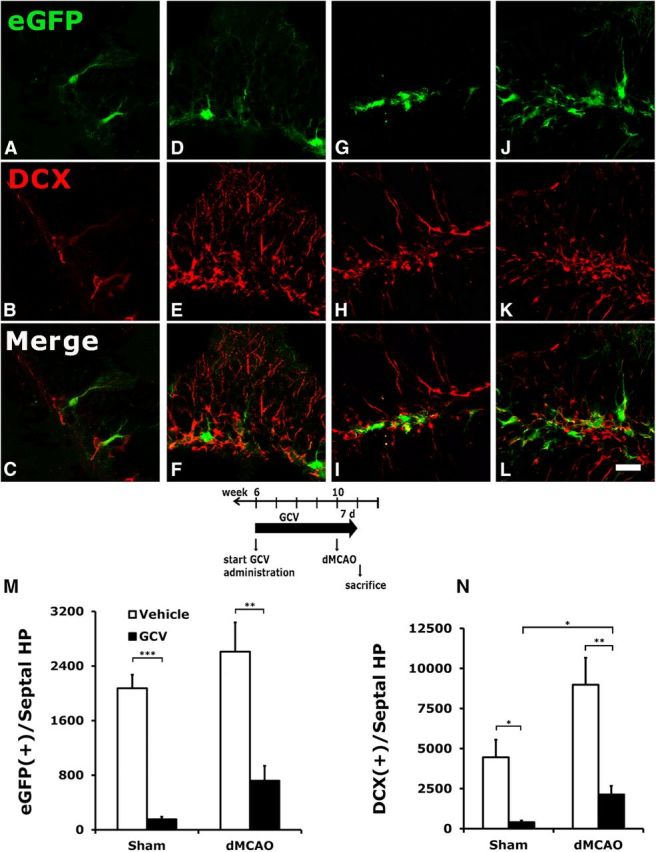
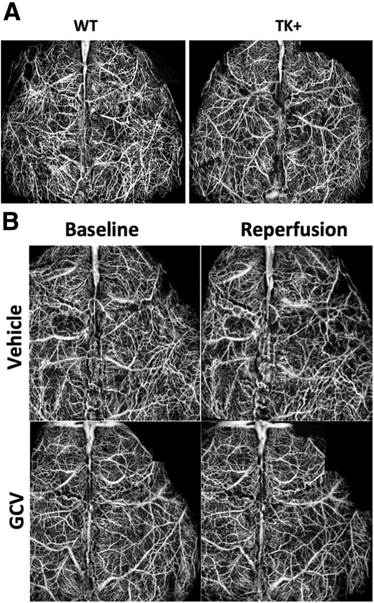
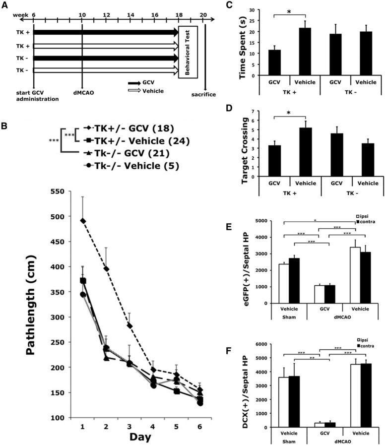
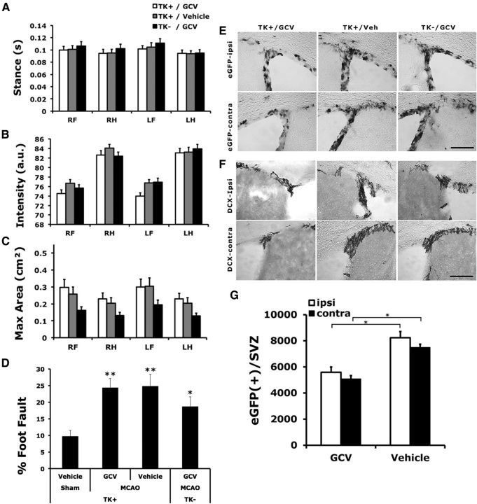
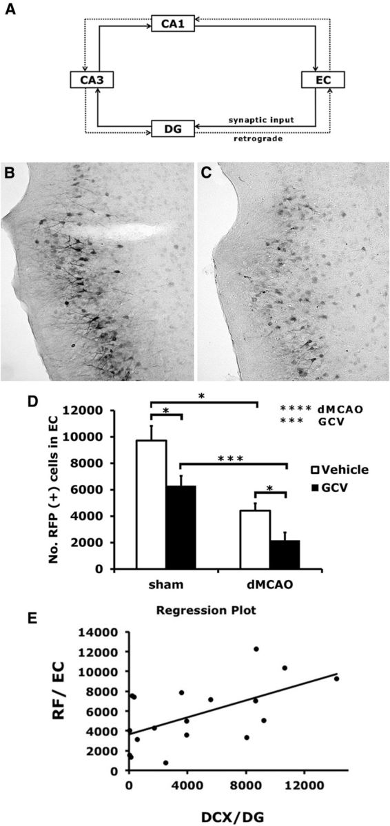
The causal relationship between neurogenesis and the recovery of poststroke cognitive function has not been properly explored. The current study aimed to determine whether depleting neuroprogenitor cells (NPCs) affects poststroke functional outcome in nestin-δ-HSV-TK-EGFP transgenic mice, in which the expression of a truncated viral thymidine kinase gene and EGFP was restricted to nestin-expressing NPCs. Ganciclovir (GCV; 200 mg/kg/d) or saline was continuously administered via osmotic pumps in mice for 4 weeks before the induction of experimental stroke. Both baseline and stroke-induced type 1 and type 2 NPCs were conditionally ablated. GCV eliminated NPCs in a duration-dependent fashion, but it did not attenuate the genesis of astroglia or oligodendrocytes in the peri-infarct cortex, nor did it affect infarct size or cerebral blood reperfusion after stroke. Transgenic stroke mice given GCV displayed impaired spatial learning and memory in the Barnes maze test compared with saline control or wild-type stroke mice given GCV, suggesting a contributing role of stroke-induced neurogenesis in the recovery of cognitive function. However, there was no significant difference in poststroke motor function between transgenic mice treated with GCV and those treated with vehicle, despite a significant ablation of NPCs in the subventricular zone of the former. Furthermore, nestin-δ-HSV-TK-EGFP mice treated with GCV had fewer retrogradely labeled neurons in the entorhinal cortex (EC) when injected with the polysynaptic viral marker PRV614 in the dentate gyrus (DG), suggesting that there might be reduced synaptic connectivity between the DG and EC following ablation of NPCs, which may contribute to impaired poststroke memory function.


Figures

Figure 1.
Figure 1.
Conditional ablation of hippocampal NPCs by GCV in TK+ mice subjected to stroke or sham surgery. A–L, Vehicle or GCV was administered to TK+ mice for 4 weeks before stroke or sham surgery, and continued for 7 d after surgery until the mice were killed. Type 1 (EGFP-immunoreactive) and type 2b (DCX-immunoreactive) progenitors were significantly reduced following GCV treatment in mice who had undergone either sham surgery (A–C) or a dMCAO procedure (G–I) compared with those treated with vehicle (D–F and J–L, respectively). M, N, Although type 1 NPCs in the DG showed a tendency to increase (M) and type 2 cells had a significant increase (N) after stroke, they were still much reduced compared with the corresponding groups in the vehicle-treated mice. GCV also reduced the degree of dendritic arborization in the type 1 NPCs in the DG (G), compared with the elaborate radial process observed in the vehicle-treated EGFP cells (J). *p < 0.05; **p < 0.01, ***p < 0.005. Scale bar, 25 μm. N = 3–4/group.
Figure 2.
Figure 2.
Following dMCAO, TK+ mice continued to receive GCV for 0, 3, 7, 14, or 28 d and were killed 28 d after stroke. A, Time lines of surgery, GCV treatment, and survival period. B, C, The numbers of EGFP-expressing (B) and DCX-expressing (C) cells in the septal hippocampus are inversely correlated with the duration of GCV treatment. Early withdrawal of GCV following dMCAO did not further significantly reduce the number of NPCs compared with mice that received GCV before stroke. However, groups that continued to receive GCV for 14 and 28 d, and significantly fewer type 1 NPCs remained in both the ipsilateral and contralateral DG. Type 2 NPCs were affected similarly. Significant statistical results are labeled above the bar of a specific duration when compared with the 0 d time point of each hemisphere. Additional significant statistics are indicated between time points. *p < 0.05; **p < 0.01; ***p < 0.005. N = 5–9/group.
Figure 3.
Figure 3.
Stroke induced the proliferation of NPCs in the DG and glial cells in the peri-infarct cortex in the presence of GCV. Following dMCAO, TK+ mice continued to receive GCV for 7 d and were killed after 28 d. A–D, BrdU was given during days 4–7 after stroke. GCV did not affect dividing reactive astrocytes (A), oligo-2-expressing cells (B), or oligodendrocyte progenitors (C) in the peri-infarct cortex, or Iba-1-expressing microglia in the infarct core (D). E, F, Few stroke-induced newly born NPCs in the DG survived and expressed either type 1 (E) or type 2 (F) cell markers despite the continued presence of GCV. G, A few newly divided NPCs differentiated into NeuN-expressing mature neurons and migrated into the granule cell layer. H, However, colocalization between BrdU and GFAP was rarely detected in the DG. I, J, Percentages of cell type-specific markers colocalized with BrdU in the periinfarct cortex or infarct core (I) and DG (J). Scale bars, 25 μm. N = 7/group.
Figure 4.
Figure 4.
Distal occlusion of MCA does not induce injury in the hippocampus. A–H, Fluoro-Jade C was used to label degenerating neurons in mice treated with vehicle (A–D) or GCV (E–H) at 4 d after dMCAO. There was no Fluoro-Jade C staining in the ipsilateral (C, G) or contralateral (D, H) hippocampus, indicating no sign of injury. However, robust Fluoro-Jade C labeling was seen in the peri-infarct region ipsilateral to ischemic stroke in both groups (A, B, E, F). B and F are 40× magnification of A and E. i, Ischemic infarct; CTX, cortex; cc, corpus callosum; DG, dentate gyrus. Scale bars: A, E, 100 μm; B, E, 25 μm; C, G, 100 μm; D, H, 200 μm. N = 4–5/group.
Figure 5.
Figure 5.
Conditional ablation of NPCs does not affect functional blood flow during reperfusion. A, Representative OCT images revealed no significant difference in baseline functional vascular network between wild-type and TK transgenic mice, suggesting a lack of transgene effect on the development of cerebrovasculature. B, There was also no significant difference in cerebral blood flow in the ipsilesional hemisphere during 1 h after reperfusion between TK transgenic mice treated with vehicle and those with GCV. N = 3–6/group.
Figure 6.
Figure 6.
Conditional ablation of NPCs impaired poststroke spatial learning and memory function in the Barnes maze test. A, Time lines of surgery and drug treatment. B, Chronic treatment of GCV in TK+ mice subjected to stroke worsened the acquisition rate over 6 d in the Barnes maze test compared with vehicle treatment (***p < 0.005) or compared with ischemic TK mice that do not carry the δ-HSV-TK transgene while being treated with GCV (**p < 0.01). Neither GCV treatment of wild-type mice with stroke (TK GCV vs TK vehicle: p > 0.78) nor transgene in the absence of GCV (TK+ Vehicle vs TK vehicle: p > 0.92) significantly affected the performance of stroke mice in the Barnes maze test. C, D, Stroke mice with reduced neurogenesis also exhibited impairment in memory recall/retention, as evidenced by reduced time spent in searching the target zone (C) and reduced frequency of crossing (D). E, F, Corroborating evidence indicated that chronic treatment of GCV significantly depleted the number of type 1 and type 2 NPCs in mice subjected to dMCAO, respectively. *p < 0.05; **p < 0.01; ***p < 0.005. Group sizes are as indicated.
Figure 7.
Figure 7.
Conditional ablation of NPCs did not significantly affect the recovery of poststroke motor function. A–D, There was no significant difference in gait function perimeters (intensity, maximum contact area, and stance; A–C) or the extent of foot fault when traversing an elevated horizontal ladder (D) among TK+ stroke mice treated with GCV, TK+ stroke mice treated with vehicle, and TK mice stroke treated with GCV, compared with TK+ vehicle-treated, sham-operated mice. Regardless of genotype or treatment, stroke mice stepped through the ladder more often than the sham-operated mice. E, F, Representative photomicrographs showing the expression of EGFP (E) and DCX (F) in groups as indicated. G, There was a significant reduction in the type 1 NPCs in the SVZ in the TK+ mice treated with GCV compared with those treated with vehicle. Scale bars, 100 μm. Comparison with the sham group: *p < 0.05; **p < 0.01. N = 18–24/group.
Figure 8.
Figure 8.
Conditional ablation of NPCs reduced synaptic connectivity in the perforant pathway. A, Trisynaptic circuits of the hippocampal network are shown in the diagram. Retrograde synaptic tracer PRV614 was injected into the DG at 4 weeks after stroke or sham surgery. B, C, Representative images of cells immunopositive for RFP from TK+ mice treated with vehicle (B) and with GCV (C), and subjected to stroke. D, Retrograde spread of the virus from the DG to EC was quantified as shown. Conditional ablation of NPCs by GCV as well as stroke both reduced the spread of PRV614 into EC. E, Regression analysis suggested that there was a positive correlation between the number of DCX-positive cells in the DG and PRV614 infectivity in the EC (p < 0.05). Y = 3639.6 + 0.4X; R2 = 0.34. N = 4–6/group.

References

    1. Aimone JB, Deng W, Gage FH. Resolving new memories: a critical look at the dentate gyrus, adult neurogenesis, and pattern separation. Neuron. 2011;70:589–596. doi: 10.1016/j.neuron.2011.05.010. - DOI - PMC - PubMed
    1. An L, Qin J, Wang RK. Ultrahigh sensitive optical microangiography for in vivo imaging of microcirculations within human skin tissue beds. Opt Express. 2010;18:8220–8228. doi: 10.1364/OE.18.008220. - DOI - PMC - PubMed
    1. Arruda-Carvalho M, Sakaguchi M, Akers KG, Josselyn SA, Frankland PW. Posttraining ablation of adult-generated neurons degrades previously acquired memories. J Neurosci. 2011;31:15113–15127. doi: 10.1523/JNEUROSCI.3432-11.2011. - DOI - PMC - PubMed
    1. Arvidsson A, Collin T, Kirik D, Kokaia Z, Lindvall O. Neuronal replacement from endogenous precursors in the adult brain after stroke. Nat Med. 2002;8:963–970. doi: 10.1038/nm747. - DOI - PubMed
    1. Bragin A, Jandó G, Nádasdy Z, Hetke J, Wise K, Buzsáki G. Gamma (40–100 Hz) oscillation in the hippocampus of the behaving rat. J Neurosci. 1995;15:47–60. - PMC - PubMed

Publication types