. 2013 Oct 30;33(44):17458-68.
doi: 10.1523/JNEUROSCI.1380-13.2013.

Receptor interacting protein kinase-mediated necrosis contributes to cone and rod photoreceptor degeneration in the retina lacking interphotoreceptor retinoid-binding protein

Affiliations

Receptor interacting protein kinase-mediated necrosis contributes to cone and rod photoreceptor degeneration in the retina lacking interphotoreceptor retinoid-binding protein

Kota Sato et al. J Neurosci. .

Abstract

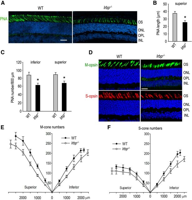
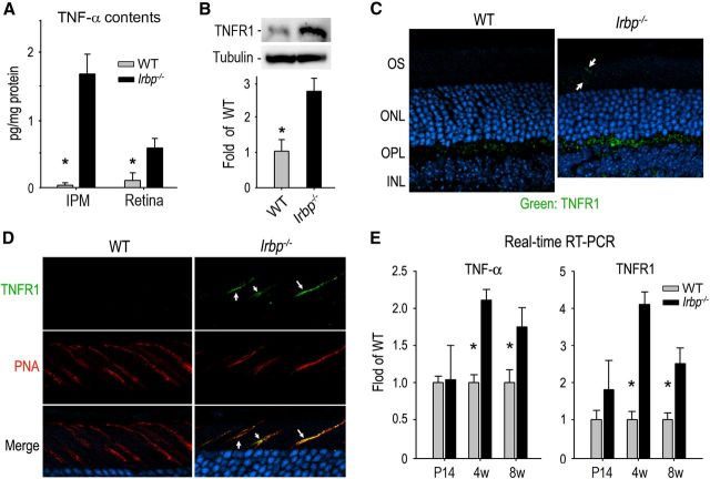
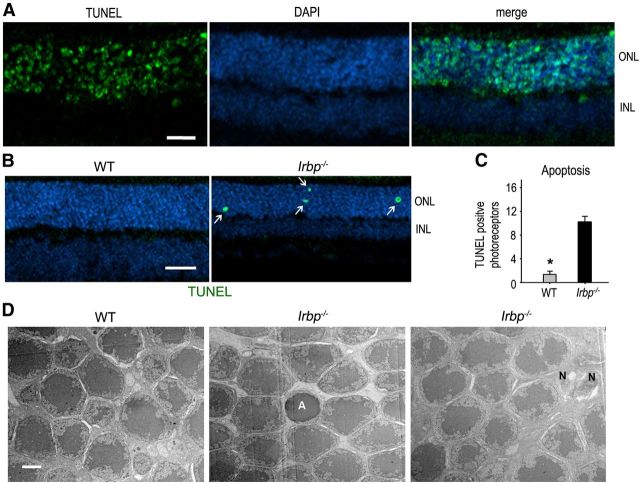
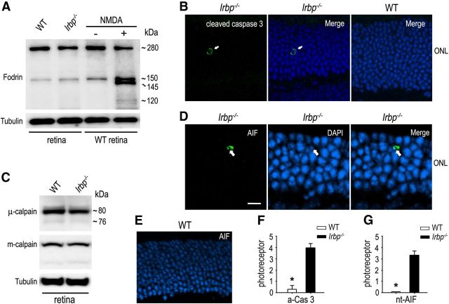
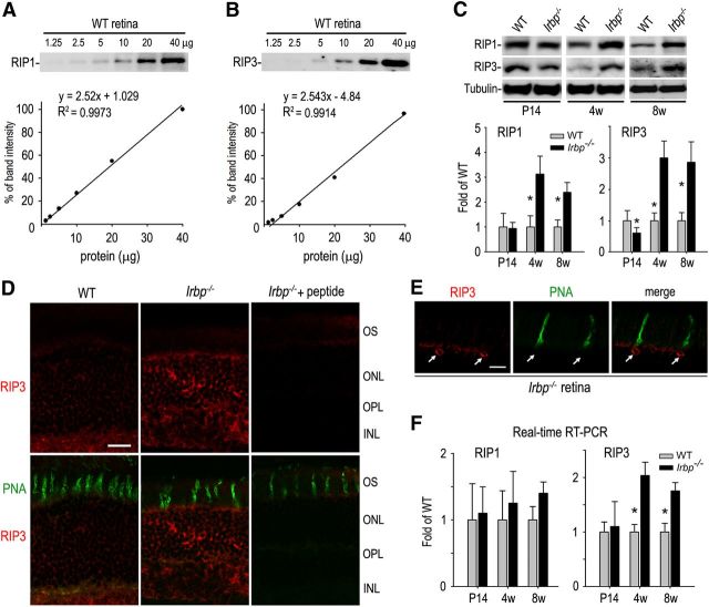
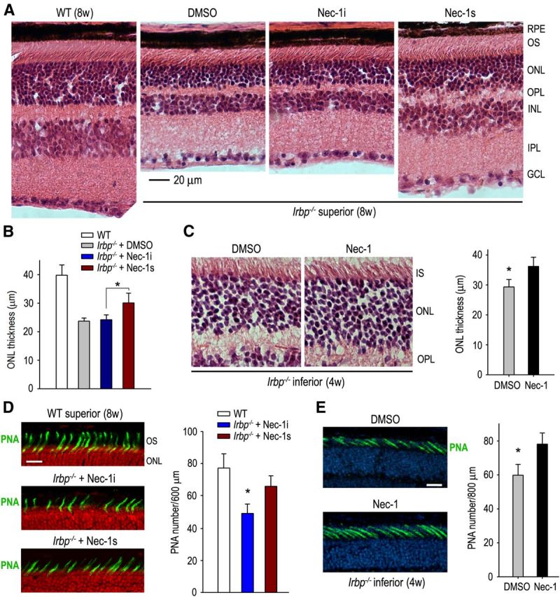
Interphotoreceptor retinoid-binding protein (IRBP) secreted by photoreceptors plays a pivotal role in photoreceptor survival with an unknown mechanism. A mutation in the human IRBP has been linked to retinitis pigmentosa, a progressive retinal degenerative disease. Mice lacking IRBP display severe early and progressive photoreceptor degeneration. However, the signaling pathway(s) leading to photoreceptor death in IRBP-deficient mice remains poorly understood. Here, we show that amounts of tumor necrosis factor-α (TNF-α) in the interphotoreceptor matrix and retinas of Irbp(-/-) mice were increased more than 10-fold and fivefold, respectively, compared with those in wild-type mice. Moreover, TNF-α receptor 1, an important membrane death receptor that mediates both programmed apoptosis and necrosis, was also significantly increased in Irbp(-/-) retina, and was colocalized with peanut agglutinin to the Irbp(-/-) cone outer segments. Although these death signaling proteins were increased, the caspase-dependent and independent apoptotic pathways were mildly activated in the Irbp(-/-) retinas, suggesting that other cell death mechanism(s) also contributes to the extensive photoreceptor degeneration in Irbp(-/-) retina. We found that receptor interacting protein 1 and 3 (RIP1 and RIP3) kinases, the intracellular key mediators of TNF-induced cellular necrosis, were elevated at least threefold in the Irbp(-/-) retinas. Moreover, pharmacological inhibition of RIP1 kinase significantly prevented cone and rod photoreceptor degeneration in Irbp(-/-) mice. These results reveal that RIP kinase-mediated necrosis strongly contributes to cone and rod degeneration in Irbp(-/-) mice, implicating the TNF-RIP pathway as a potential therapeutic target to prevent or delay photoreceptor degeneration in patients with retinitis pigmentosa caused by IRBP mutation.


Figures

Figure 1.
Figure 1.
Cone and rod degeneration in Irbp−/− retinas. A, Cone matrix sheathes in the superior retinas of 4-week-old WT and Irbp−/− mice were stained with fluorescein-tagged PNA. Nuclei were counterstained with DAPI (blue). OS of photoreceptors, ONL, OPL, and INL are indicated. Scale bar, 20 μm. B, Histograms showing length of PNA-positive cone matrix sheathes in the central retinas of WT and Irbp−/− mice. C, Histograms showing counts of PNA-positive cone matrix sheathes in 800 μm width regions of the inferior or superior retinas of WT and Irbp−/− mice. Asterisks indicate statistically significant differences between WT and mutant retinas (p < 0.01). Error bars denote SD (n = 4). D, Representative immunohistochemistry showing M-opsin (green)-positive cone OS in the superior and S-opsin (red)-positive cone OS in the inferior retinas of 4-week-old WT and Irbp−/− mice. Scale bar, 20 μm. E, F, Counts of cone OS positive for M-opsin or S-opsin. Numbers on the x-axis indicate distance from optic nerve head. Error bars denote SD (n = 3).
Figure 2.
Figure 2.
Upregulation of TNF-α and TNFR1 in Irbp−/− retina. A, Amounts of TNF-α in 4-week-old (4w) WT and Irbp−/− IPM and retinas were determined by the enzyme-linked immuno sorbent assay. Asterisks indicate significant differences between WT and mutant mice (p < 0.01). Error bars denote SD (n = 4). B, Immunoblot analysis of TNFR1 in 4w-old WT and Irbp−/− retinas. Relative expression levels of TNFR1 in WT and Irbp−/− retinas were normalized to tubulin levels in three independent experiments and shown in the histogram. C, Representative immunohistochemistry showing upregulation of TNFR1 in Irbp−/− retina. Arrows indicate photoreceptor OS positive for TNFR1. D, Double staining of TNFR1 (green) and rhodamine-tagged PNA (red) in WT and Irbp−/− retinal sections. Arrows indicate photoreceptor OS positive for both TNFR1 and PNA staining. E, Relative expression levels of TNF-α and TNFR1 mRNAs in WT and Irbp−/− retinas at the indicated ages were determined by quantitative RT-PCR and were normalized to 18S rRNA levels. Error bars designate SD (n = 4).
Figure 3.
Figure 3.
Apoptosis of photoreceptors in Irbp−/− retina. A, Photoreceptor apoptosis (green label) in WT mice was induced with an injection of NMU, and was detected by TUNEL assay as a positive control. Nuclei were counterstained with DAPI. B, Retinal sections from 4-week-old WT and Irbp−/− mice were analyzed by TUNEL assay to detect photoreceptor apoptosis. Arrows indicate TUNEL-positive photoreceptors. Scale bars, 20 μm. C, Histogram showing the average counts of TUNEL-positive photoreceptors in a whole retinal section from 4-week-old WT and Irbp−/− mice. Asterisks indicate significant differences between WT and Irbp−/− mice (p < 0.001). Error bars designate SD (n = 4). D, TEM photomicrographs of photoreceptors in 4-week-old WT and Irbp−/− retinas. A and N in the images indicate apoptotic and necrotic cells, respectively. Scale bar, 2 μm.
Figure 4.
Figure 4.
Caspase-dependent and -independent apoptotic pathways were activated in a few Irbp−/− photoreceptors. A, Immunoblot analysis of α-fodrin in 4-week-old WT and Irbp−/− retinas as well as in WT retinas treated (+) or untreated (−) with NMDA. Caspase-3-cleaved 120 and 150 kDa fragments of α-fodrin are indicated. Tubulin was detected as a loading control. B, Immunohistochemistry showing the presence of active caspase-3 (green, arrow) in a nucleus of Irbp−/− photoreceptor, but not WT photoreceptor. Nuclei were counterstained with DAPI (blue). C, Immunoblot analysis of m-calpains and μ-calpains in WT and Irbp−/− retinas. The 80 kDa proenzyme and 76 kDa active form of calpains are indicated. D, Representative immunohistochemistry showing translocation of AIF into an Irbp−/− photoreceptor nucleus (green, arrow). Scale bar, 5 μm. E, Immunohistochemistry showing the absence of AIF-positive nucleus in WT photoreceptors. F, G, Histograms showing the average counts of photoreceptors positive for active caspase 3 (a-Cas3) or nuclear-translocated AIF (nt-AIF) in a whole retinal section from WT and Irbp−/− mice. Asterisks indicate significant differences between WT and Irbp−/− mice (p < 0.001). Error bars designate SD (n = 4).
Figure 5.
Figure 5.
Upregulation of RIP1 and RIP3 kinases in Irbp−/− retina. A, B, Calibration curves showing linearity of band intensities of RIP1 (A) and RIP3 (B) versus retinal protein amounts over the ranges 1.25–40 μg. The densitometry data from 40 μg of protein was set as 100% signal intensity on the y-axis. C, Immunoblot analysis for RIP1 and RIP3 in retinas of WT and Irbp−/− mice at the indicated ages. Expression levels of RIP1 and RIP3 in Irbp−/− retinas were normalized to tubulin, and shown as fold of those in WT retinas. Asterisks indicate significant differences between WT and Irbp−/− mice (p < 0.01). Error bars designate SD (n = 4). D, Representative immunohistochemistry showing increased expression levels of RIP3 in the ONL, OPL, and INL of Irbp−/− retinal sections stained with a RIP3 antibody in the presence (+ peptide) or absence of immunogenic peptides. Cone matrix sheathes were stained with PNA (green). Scale bar, 10 μm. E, High-magnification images of double staining for RIP3 (red) and PNA (green). Arrows indicate expression of RIP3 in cones. Scale bar, 5 μm. F, Expression levels of RIP1 and RIP3 mRNAs in Irbp−/− retinas at the indicated ages were determined by quantitative RT-PCR, normalized to 18S rRNA, and expressed as fold of their expression levels in WT retinas. Asterisks indicate significant differences between WT and Irbp−/− (p < 0.01). Error bars indicate SD (n = 4).
Figure 6.
Figure 6.
Protection of cone and rod photoreceptors by RIP1 inhibitors in Irbp−/− mice. A, H&E staining of retinal sections from 8-week-old (8w) WT and Irbp−/− mice treated with DMSO, Nec-1i, or Nec-1s. GCL, Ganglion cell layer. B, Histogram showing the average thickness of the ONL in superior retinas from 8w-old WT and Irbp−/− mice treated with the indicated reagents. Asterisks indicate statistically significant differences between mice treated with Nec-1i or Nec-1s (p < 0.05). Error bars denote SD (n = 6). C, H&E staining of retinal sections of 4w-old Irbp−/− mice treated with DMSO or Nec-1. The average thickness of the ONL in the inferior retinas of the mice is shown in the histogram. Asterisks indicate significant differences between the two treatments (p < 0.05). Error bars denote SD (n = 6). IS, Inner segment of photoreceptor. D, PNA staining (green) of the superior retinas of 8w-old WT and Irbp−/− mice treated with Nec-1i or Nec-1s. Nuclei were counterstained with propidium iodide (red). Numbers of PNA-positive cone matrix sheathes in a 600 μm width region of the superior retinas are shown in the histogram. Scale bar, 20 μm. E, PNA staining of the inferior retinas of 4w-old Irbp−/− mice treated with DMSO or Nec-1. Nuclei were counterstained with DAPI (blue). Numbers of PNA-positive cone sheathes in 800 μm width region of the mice inferior retinas are shown in the histogram. Scale bar, 10 μm.

Similar articles

Cited by

References

    1. Ashkenazi A, Dixit VM. Death receptors: signaling and modulation. Science. 1998;281:1305–1308. doi: 10.1126/science.281.5381.1305. - DOI - PubMed
    1. Bazan NG, Reddy TS, Redmond TM, Wiggert B, Chader GJ. Endogenous fatty acids are covalently and noncovalently bound to interphotoreceptor retinoid-binding protein in the monkey retina. J Biol Chem. 1985;260:13677–13680. - PubMed
    1. Black RA, Rauch CT, Kozlosky CJ, Peschon JJ, Slack JL, Wolfson MF, Castner BJ, Stocking KL, Reddy P, Srinivasan S, Nelson N, Boiani N, Schooley KA, Gerhart M, Davis R, Fitzner JN, Johnson RS, Paxton RJ, March CJ, Cerretti DP. A metalloproteinase disintegrin that releases tumour-necrosis factor-alpha from cells. Nature. 1997;385:729–733. doi: 10.1038/385729a0. - DOI - PubMed
    1. Borst DE, Redmond TM, Elser JE, Gonda MA, Wiggert B, Chader GJ, Nickerson JM. Interphotoreceptor retinoid-binding protein. Gene characterization, protein repeat structure, and its evolution. J Biol Chem. 1989;264:1115–1123. - PubMed
    1. Bridges CD, Alvarez RA, Fong SL, Gonzalez-Fernandez F, Lam DM, Liou GI. Visual cycle in the mammalian eye. Retinoid-binding proteins and the distribution of 11-cis retinoids. Vision Res. 1984;24:1581–1594. doi: 10.1016/0042-6989(84)90316-X. - DOI - PubMed

Publication types

MeSH terms

Substances