. 2013 Oct 30;33(44):17527-37.
doi: 10.1523/JNEUROSCI.0857-13.2013.

CXC chemokine receptor 7 (CXCR7) affects the migration of GnRH neurons by regulating CXCL12 availability

Affiliations

CXC chemokine receptor 7 (CXCR7) affects the migration of GnRH neurons by regulating CXCL12 availability

Fani Memi et al. J Neurosci. .

Abstract

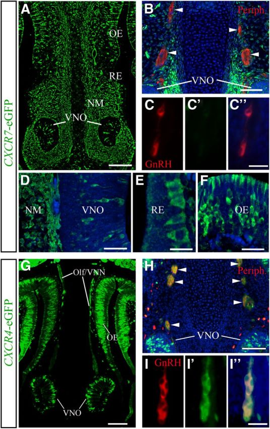
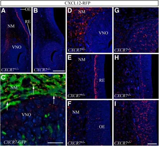
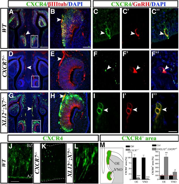
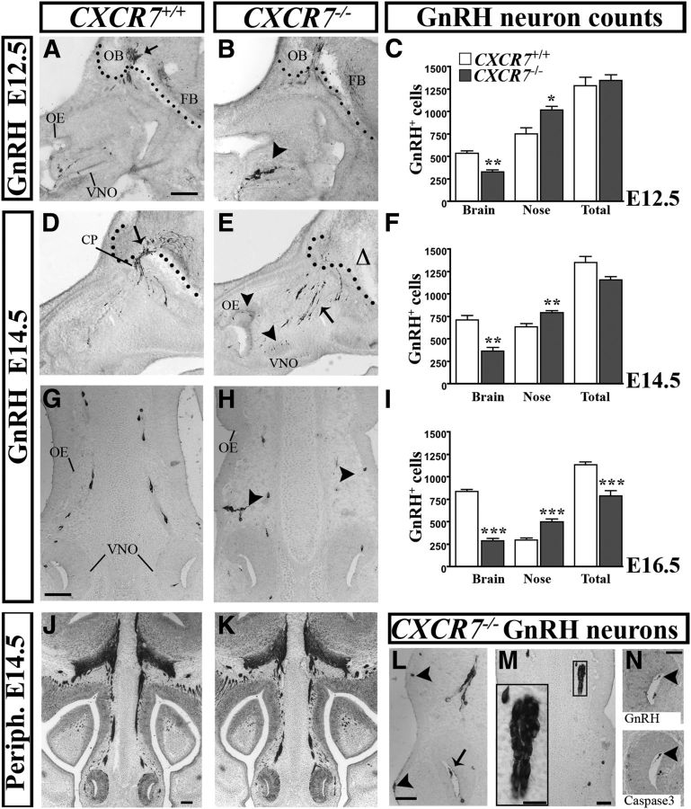
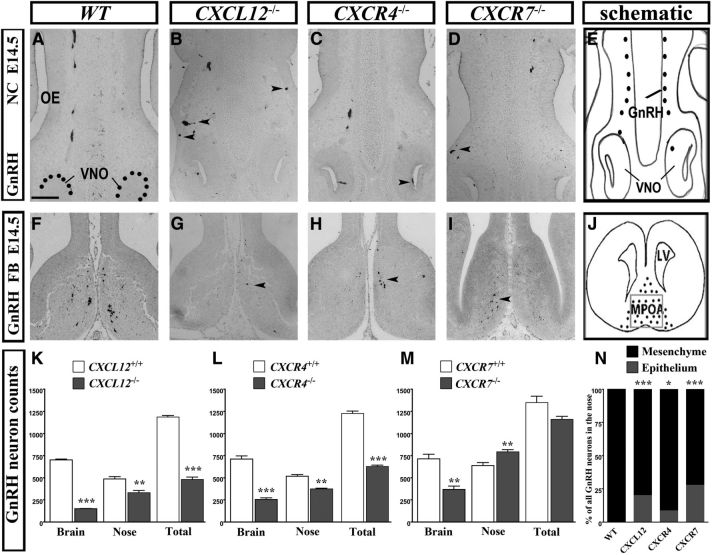
Gonadotropin-releasing hormone (GnRH) neurons are neuroendocrine cells, located in the hypothalamus, that play an essential role in mammalian reproduction. These neurons originate in the nasal placode and migrate during embryonic development, in association with olfactory/vomeronasal nerves, first in the nose, then through the cribriform plate to enter the forebrain, before settling in the hypothalamus. One of the molecules required for their early migration in the nose is the chemokine CXCL12, which is expressed in the embryonic nasal mesenchyme in an increasing ventral to dorsal gradient, presumably guiding GnRH neurons toward the forebrain. Mice lacking CXCR4, the receptor for CXCL12, exhibit defective GnRH cell movement and a significant reduction in their number, suggesting that CXCL12/CXCR4 signaling is important in the migration and survival of these neurons. Here, we investigated the role of the more recently identified second CXCL12 receptor, CXCR7, in GnRH neuron development. We demonstrate that CXCR7 is expressed along the migratory path of GnRH neurons in the nasal cavity and, although not expressed by GnRH neurons, it affects their migration as indicated by the ectopic accumulation of these cells in the nasal compartment in CXCR7(-/-) mice. Absence of CXCR7 caused abnormal accumulation of CXCL12-RFP at CXCR4-positive sites in the nasal area of CXCL12-RFP-transgenic mice and excessive CXCL12-dependent intracellular clustering of CXCR4 in GnRH neurons, suggesting internalization. These findings imply that CXCR7 regulates CXCL12 availability by acting as a scavenger along the migratory path of GnRH neurons and, thus, influences the migration of these cells in a noncell-autonomous manner.


Figures

Figure 1.
Figure 1.
Expression of CXCL12 and its receptors, CXCR7 and CXCR4, in the nasal area of E14.5 mice as revealed by in situ hybridization. A, Low-magnification image of a coronal head section at the level of the nose reveals CXCR7 expression in the VNO, NM, RE, and OE. A′, A″, Higher magnification views show that CXCR7 is predominantly expressed in the apical zone of the sensory epithelium of VNO (arrow) and OE. B, CXCL12 is expressed in the VNO and NM. A′, B′, CXCL12 and CXCR7 overlap in the apical zone (AZ) of the VNO and the NM dorsomedial to the VNO. B″, Higher magnification of the OE area where CXCL12 is highly expressed in the NM and moderately expressed in the dorsal part of the sensory epithelium. Note that most of the OE is CXCL12 negative. C, CXCR4 is highly expressed in the sensory epithelium of VNO and OE and sparsely expressed in the RE. Presumed migrating GnRH neurons are CXCR4 positive (arrows). C′, C″, Higher magnification images illustrate predominant CXCR4 expression in the basal zone (BZ) of VNO (C′) and OE (C″). D, D′, In situ hybridization for CXCL12 and immunohistochemistry for GnRH illustrate migrating GnRH neurons (arrows) on CXCL12 expressing substrate in the NM. D′, D″, These neurons are shown in higher magnification in the area of the VNO (D′) and OE (D″). Scale bars: (in A) A–D, 200 μm; in (A′) A′–D″, 100 μm. CB, cavernous body; CC, cartilaginous capsule.
Figure 2.
Figure 2.
GnRH neurons and their axonal scaffold do not express CXCR7, but do express CXCR4. A, G, Coronal head sections of E14.5 transgenic CXCR7- and CXCR4-eGFP mice reveal the relative expression patterns of CXCR7 and CXCR4 in the nasal area. B, H, Olf/VNN axons (arrowheads) used by migrating GnRH neurons and stained for peripherin (red) are CXCR7 negative (B), but CXCR4 positive (H). C, I, Dual immunofluorescence for GnRH and eGFP demonstrates that GnRH neurons are eGFP negative in CXCR7-eGFP mice (C–C″) and eGFP positive in CXCR4-eGFP mice (I–I″) indicating that GnRH neurons express CXCR4, but not CXCR7. D–F, High-magnification images reveal CXCR7-eGFP in the VNO area (D), RE (E), and OE (F) of E14.5 mice. Specimens shown in B–F, H, and I were counterstained with DAPI. A–I, All images were acquired by confocal microscopy. Scale bars: A, 140 μm; B, 50 μm; (in C″) C–C″, 50 μm; D, 30 μm; E, 14 μm; F, 30 μm; G, 100 μm; H, 100 μm; (in I″) I–I″, 50 μm. Olf/VNN, olfactory/vomeronasal nerve; Periph, peripherin.
Figure 3.
Figure 3.
CXCR7 regulates spatial distribution of CXCL12-RFP along the migration route of GnRH neurons. A–I, Immunostaining for RFP in coronal sections of the nasal area of E14.5 transgenic mice expressing CXCL12-RFP fusion protein. A, B, Low-magnification confocal images reveal strongly decreased RFP signal in the NM and RE and increased RFP signal in the VNO and OE of a CXCL12-RFP;CXCR7−/− embryo (B) as compared with a CXCL12-RFP;CXCR7+/− littermate (A). D–I, High-magnification confocal views of VNO and adjacent NM (D, G), RE (E, H), and OE (F, I). C, High-magnification confocal view of a dual immunofluorescence for RFP and eGFP in a CXCL12-RFP;CXCR7-eGFP bitransgenic mouse shows RFP signal in eGFP-positive mesenchymal cells (arrows). Scale bars: (in B) A, B, 50 μm; C, 14 μm; (in I) D–I, 14 μm.
Figure 4.
Figure 4.
Subcellular redistribution and loss of CXCR4 in the nasal area of CXCR7−/− mice. A–M, Immunostained frontal sections of E14.5 mice. A, B, Double labeling for CXCR4 (green) and βIII tubulin (red) shows that in a WT animal, CXCR4 is localized in axons (arrowheads) and in cells in the VNO (B). D, E, CXCR4 is not detectable in axons (arrowheads) and shows a punctate/internalized pattern in the VNO of a CXCR7−/− animal (E). G, H, The loss of CXCR4 observed after CXCR7 ablation (D, E) is reversed in a CXCL12−/−;CXCR7−/− animal (XL12−/−;X7−/−). C, F, I, Similar changes in CXCR4 localization were noted in GnRH neurons. For example, in one of these neurons of a WT animal (C–C″, arrowhead), CXCR4 is clearly expressed on the cell membrane. However, in a GnRH neuron of CXCR7−/− mutant, the receptor is localized in the cytoplasm and not detected on the plasma membrane (F–F″, arrowhead). In the absence of CXCL12, CXCR4 is upregulated on the cell membrane and intracellular clusters of CXCR4 are not observed in CXCR7-deficient GnRH neurons (I–I″, arrowhead). J–L, High-magnification views demonstrate altered subcellular distribution of CXCR4 in the OE of a CXCR7−/− mutant (K) and a CXCL12−/−;CXCR7−/− double mutant (L) as compared with a WT control (J). A–L, All images were acquired by confocal microscopy. M, Quantification of the CXCR4-immunoreactive area in the OE and VNO of CXCR7−/− and CXCL12−/−;CXCR7−/− mice and their control littermates (n = 4). *p < 0.05, **p < 0.01,***p < 0.001, ANOVA. Green areas in the schematic represent the regions of interest used for quantification. The nasal septum is to the left and the nasal cavity (shaded) to the right. Scale bars: (in A) A, D, G, 200 μm; (in B) B, E, H, 50 μm; (in I″) C, F, I, 20 μm; (in J) J–L, 20 μm. WT, wild-type.
Figure 5.
Figure 5.
Migration of GnRH neurons is altered in CXCR7-null mice. Sagittal sections (A, B) through the nose at E12.5, stained for GnRH, show that cells tend to remain in the area of the VNO (B, arrowhead) in CXCR7−/− mice whereas, in the corresponding controls, they appear to cross the cribriform plate (arrow) and migrate toward the forebrain (FB). Black dots separate the nasal area from the brain. D, E, A similar picture is evident at 14.5, with most GnRH neurons still migrating in the nasal area in CXCR7-null mice (E, arrow). Additionally, some cells are still present in the VNO and, abnormally, in the OE (E, arrowheads). The relative absence of GnRH neurons in the forebrain is indicated with a triangle. G, H, Coronal sections stained for GnRH reveal that, in CXCR7−/− mice, migrating cells deviate from their normal chain-like formation (G) and, instead, appear in the OE and RE (arrowheads in H and L). Further, they form abnormal multicellular clusters in the NM (rectangle in M and at higher magnification in the inset in M) and, sometimes, in the lumen of the VNO (L, arrow). N, GnRH cells in the lumen of the VNO (arrowhead) show cleaved caspace-3 in a contiguous section, suggesting they are destined to die. J, K, However, guiding axons stained for peripherin appear similar in both genotypes. C, F, I, Compartmental quantification of GnRH cells in the three ages examined showed significant increase of neurons in the nose and a concomitant decrease in the brain of CXCR7−/− mice compared with WT. Moreover, while the total number of cells was similar in the two groups at E12.5 and E14.5, it declined in the KO mice at E16.5 (I). *p < 0.05, **p < 0.01, ***p < 0.001; Scale bars: (in A) A, B, 150 μm; D, E, G, H, L, M, 50 μm; (in J) J, K, 500 μm; N, 30 μm. CP, cribriform plate; OB, olfactory bulb.
Figure 6.
Figure 6.
GnRH neuron migration in CXCL12−/−, CXCR4−/−, and CXCR7−/− mice at E14.5. A–D, In the nose, GnRH neurons migrate in a chain-like formation in WT animals but, in KOs, they form clusters (B, arrowheads), and are often found abnormally in the lumen of the VNO (C, arrowhead) or in the epithelium (D, arrowhead). F–I, GnRH staining of coronal sections at the level of the MPOA shows the distribution of these cells in WT animals (F) and the relative reduction in numbers in the CXCL12−/− (G), CXCR4−/− (H), and CXCR7−/− (I) mice. K–M, Compartmental quantification of GnRH neurons shows an overall decrease in CXCL12−/− and CXCR4−/−, but not in CXCR7−/− animals, and a significant reduction in both nose and brain when compared with control animals. N, Spread/distribution analysis of GnRH neurons in the nasal area, divided in two compartments, epithelium and NM, show that 22% of all cells in the nasal area are abnormally localized in the epithelium of CXCL12−/− animals. The respective percentage is 28% in CXCR7−/− and 8.5% in CXCR4−/− mice. No GnRH neurons are found in the epithelium in control animals. E, J, Schematics show the nasal compartment (NC) and the medial preoptic area (MPOA) in the forebrain (FB) as photographed in A–D and F–I, respectively. K–N, *p < 0.05, **p < 0.01,***p < 0.001. Scale bars: (in A) A–D, F–I, 100 μm. FB, forebrain; LV, lateral ventricle.

Similar articles

Cited by

References

    1. Balabanian K, Lagane B, Infantino S, Chow KY, Harriague J, Moepps B, Arenzana-Seisdedos F, Thelen M, Bachelerie F. The chemokine SDF-1/CXCL12 binds to and signals through the orphan receptor RDC1 in T lymphocytes. J Biol Chem. 2005;280:35760–35766. doi: 10.1074/jbc.M508234200. - DOI - PubMed
    1. Bhattacharyya BJ, Banisadr G, Jung H, Ren D, Cronshaw DG, Zou Y, Miller RJ. The chemokine stromal cell-derived factor-1 regulates GABAergic inputs to neural progenitors in the postnatal dentate gyrus. J Neurosci. 2008;28:6720–6730. doi: 10.1523/JNEUROSCI.1677-08.2008. - DOI - PMC - PubMed
    1. Boldajipour B, Mahabaleshwar H, Kardash E, Reichman-Fried M, Blaser H, Minina S, Wilson D, Xu Q, Raz E. Control of chemokine-guided cell migration by ligand sequestration. Cell. 2008;132:463–473. doi: 10.1016/j.cell.2007.12.034. - DOI - PubMed
    1. Borrell V, Marín O. Meninges control tangential migration of hem-derived Cajal-Retzius cells via CXCL12/CXCR4 signaling. Nat Neurosci. 2006;9:1284–1293. doi: 10.1038/nn1764. - DOI - PubMed
    1. Burns JM, Summers BC, Wang Y, Melikian A, Berahovich R, Miao Z, Penfold ME, Sunshine MJ, Littman DR, Kuo CJ, Wei K, McMaster BE, Wright K, Howard MC, Schall TJ. A novel chemokine receptor for SDF-1 and I-TAC involved in cell survival, cell adhesion, and tumor development. J Exp Med. 2006;203:2201–2213. doi: 10.1084/jem.20052144. - DOI - PMC - PubMed

Publication types

LinkOut - more resources