

Machine Learning in Computer-aided Diagnosis of the Thorax and Colon in CT: A Survey
- PMID: 24174708
- PMCID: PMC3810349
- DOI: 10.1587/transinf.e96.d.772
Machine Learning in Computer-aided Diagnosis of the Thorax and Colon in CT: A Survey
Abstract
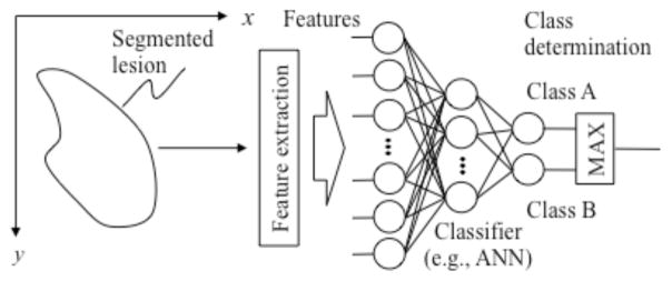
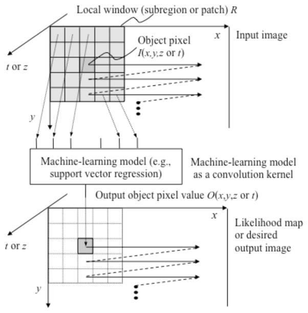
Computer-aided detection (CADe) and diagnosis (CAD) has been a rapidly growing, active area of research in medical imaging. Machine leaning (ML) plays an essential role in CAD, because objects such as lesions and organs may not be represented accurately by a simple equation; thus, medical pattern recognition essentially require "learning from examples." One of the most popular uses of ML is the classification of objects such as lesion candidates into certain classes (e.g., abnormal or normal, and lesions or non-lesions) based on input features (e.g., contrast and area) obtained from segmented lesion candidates. The task of ML is to determine "optimal" boundaries for separating classes in the multidimensional feature space which is formed by the input features. ML algorithms for classification include linear discriminant analysis (LDA), quadratic discriminant analysis (QDA), multilayer perceptrons, and support vector machines (SVM). Recently, pixel/voxel-based ML (PML) emerged in medical image processing/analysis, which uses pixel/voxel values in images directly, instead of features calculated from segmented lesions, as input information; thus, feature calculation or segmentation is not required. In this paper, ML techniques used in CAD schemes for detection and diagnosis of lung nodules in thoracic CT and for detection of polyps in CT colonography (CTC) are surveyed and reviewed.
Keywords: CT colonography; classification; colorectal polyp; computer-aided diagnosis; lung nodule; machine learning in medical imaging; pixel-based machine learning.
Figures
Similar articles
-
Pixel-based machine learning in medical imaging.Int J Biomed Imaging. 2012;2012:792079. doi: 10.1155/2012/792079. Epub 2012 Feb 28. Int J Biomed Imaging. 2012. PMID: 22481907 Free PMC article.
-
A review of computer-aided diagnosis in thoracic and colonic imaging.Quant Imaging Med Surg. 2012 Sep;2(3):163-76. doi: 10.3978/j.issn.2223-4292.2012.09.02. Quant Imaging Med Surg. 2012. PMID: 23256078 Free PMC article.
-
Improved lung nodule diagnosis accuracy using lung CT images with uncertain class.Comput Methods Programs Biomed. 2018 Aug;162:197-209. doi: 10.1016/j.cmpb.2018.05.028. Epub 2018 May 18. Comput Methods Programs Biomed. 2018. PMID: 29903487
-
Computer-aided detection for CT colonography: update 2007.Abdom Imaging. 2007 Sep-Oct;32(5):571-81. doi: 10.1007/s00261-007-9293-2. Abdom Imaging. 2007. PMID: 17690932 Review.
-
Deep learning-based CAD schemes for the detection and classification of lung nodules from CT images: A survey.J Xray Sci Technol. 2020;28(4):591-617. doi: 10.3233/XST-200660. J Xray Sci Technol. 2020. PMID: 32568165 Review.
Cited by
-
Fifty years of computer analysis in chest imaging: rule-based, machine learning, deep learning.Radiol Phys Technol. 2017 Mar;10(1):23-32. doi: 10.1007/s12194-017-0394-5. Epub 2017 Feb 16. Radiol Phys Technol. 2017. PMID: 28211015 Free PMC article. Review.
-
Machine learning for autism spectrum disorder diagnosis using structural magnetic resonance imaging: Promising but challenging.Front Neuroinform. 2022 Sep 28;16:949926. doi: 10.3389/fninf.2022.949926. eCollection 2022. Front Neuroinform. 2022. PMID: 36246393 Free PMC article. Review.
-
Overview of deep learning in medical imaging.Radiol Phys Technol. 2017 Sep;10(3):257-273. doi: 10.1007/s12194-017-0406-5. Epub 2017 Jul 8. Radiol Phys Technol. 2017. PMID: 28689314 Review.
-
Progress in Fully Automated Abdominal CT Interpretation.AJR Am J Roentgenol. 2016 Jul;207(1):67-79. doi: 10.2214/AJR.15.15996. Epub 2016 Apr 21. AJR Am J Roentgenol. 2016. PMID: 27101207 Free PMC article. Review.
-
Deep Learning in CT Images: Automated Pulmonary Nodule Detection for Subsequent Management Using Convolutional Neural Network.Cancer Manag Res. 2020 Apr 29;12:2979-2992. doi: 10.2147/CMAR.S239927. eCollection 2020. Cancer Manag Res. 2020. PMID: 32425607 Free PMC article.
References
-
- Giger ML, Suzuki K. Computer-Aided Diagnosis (CAD) In: Feng DD, editor. Biomedical Information Technology. Academic Press; 2007. pp. 359–374.
-
- Doi K. Current status and future potential of computer-aided diagnosis in medical imaging. Br J Radiol. 2005;78 (1):S3–S19. - PubMed
-
- Chan HP, Sahiner B, Helvie MA, Petrick N, Roubidoux MA, Wilson TE, Adler DD, Paramagul C, Newman JS, Sanjay-Gopal S. Improvement of radiologists’ characterization of mammographic masses by using computer-aided diagnosis: an ROC study. Radiology. 1999 Sep;212(3):817–827. - PubMed
-
- Li F, Aoyama M, Shiraishi J, Abe H, Li Q, Suzuki K, Engelmann R, Sone S, Macmahon H, Doi K. Radiologists’ performance for differentiating benign from malignant lung nodules on high-resolution CT using computer-estimated likelihood of malignancy. AJR Am J Roentgenol. 2004 Nov;183(5):1209–1215. - PubMed
-
- Li F, Arimura H, Suzuki K, Shiraishi J, Li Q, Abe H, Engelmann R, Sone S, MacMahon H, Doi K. Computer-aided detection of peripheral lung cancers missed at CT: ROC analyses without and with localization. Radiology. 2005 Nov;237(2):684–690. - PubMed
Grants and funding
LinkOut - more resources
Full Text Sources
Other Literature Sources
Miscellaneous