An unusual clinical presentation of gingival melanoacanthoma
- PMID: 24174763
- PMCID: PMC3808024
- DOI: 10.4103/0972-124X.119288
An unusual clinical presentation of gingival melanoacanthoma
Abstract
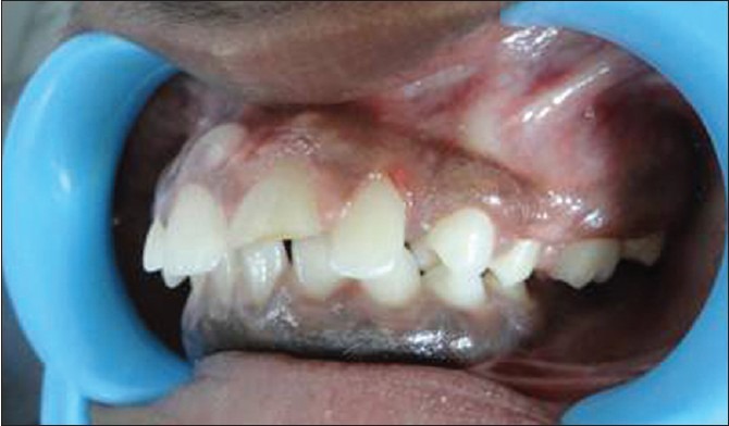
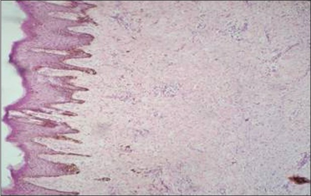
Gingival melanoacanthoma is a rare, benign pigmented lesion characterized clinically by sudden onset and rapid growth of a macular brown black lesion and histologically by acanthosis of superficial epithelium and proliferation of dendritic melanocytes. This article reports a previously undescribed case of pigmented unilateral diffuse gingival enlargement, which on histopathological examination proved to be melanoacanthoma. Intraoral examination revealed pigmented unilateral diffuse gingival enlargement in relation to second and third quadrants buccally, palatally/lingually. Based on these clinical findings, gingivectomy was performed and the excised tissue was sent for biopsy. Microscopic examination revealed acanthotic and parakeratotic surface epithelium with dendritic melanocytes distributed in basal and suprabasal layers of the epithelium. 1 year follow-up recall revealed no recurrence of lesion at the surgical sites. Our patient exhibits an unusual clinical presentation of melanoacanthoma of gingiva. Pigmented gingival overgrowth of recent origin and without any etiologic factors warrants histopathologic examination.
Keywords: Gingival enlargement; gingivectomy; melanoacanthoma.
Conflict of interest statement
Figures


References
-
- Neville BW, Damm DD, Allen CW, Bouquot JE. Epithelial pathology. In: Neville BW, Damm DD, Allen CW, Bouquot JE, editors. Oral and Maxillofacial Pathology. 3rd ed. Philadelphia: W. B. Saunders; 2009. pp. 380–2.
-
- Goode RK, Crawford BE, Callihan MD, Neville BW. Oral melanoacanthoma: Review of the literature and report of ten cases. Oral Surg Oral Med Oral Pathol. 1983;56:622–8. - PubMed
-
- Yarom N, Hirshberg A, Buchner A. Solitary and multifocal oral melanoacanthoma. Int J Dermatol. 2007;46:1232–6. - PubMed
-
- Brooks JK, Sindler AJ, Papadimitriou JC, Francis LA, Scheper MA. Multifocal melanoacanthoma of the gingiva and hard palate. J Periodontol. 2009;80:527–32. - PubMed
-
- Maize JC. Mucosal melanosis. Dermatol Clin. 1988;6:283–93. - PubMed
Publication types
LinkOut - more resources
Full Text Sources
Other Literature Sources