

Primary malignant peripheral nerve sheath tumor at unusual location
- PMID: 24174807
- PMCID: PMC3808069
- DOI: 10.4103/0976-3147.116480
Primary malignant peripheral nerve sheath tumor at unusual location
Abstract
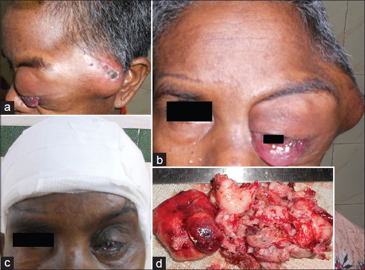
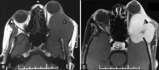
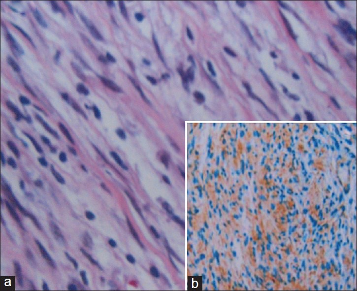
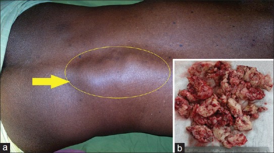
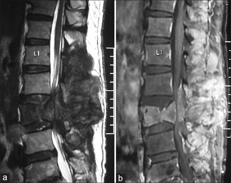
Malignant peripheral nerve sheath tumor (MPNST) is a rare soft tissue sarcoma. Most arise in association with major nerve trunks. Their most common anatomical sites are the proximal portions of the upper and lower extremities and the trunk. MPNSTs have rarely been reported in literature to occur in other unusual body parts. We review all such cases reported till now in terms of site of origin, surgical treatment, adjuvant therapy and outcome and shortly describe our experience with two of these cases. Both of our case presented with lump at unusual sites resembling neurofibroma, one at orbitotemporal area and other in the paraspinal region with characteristic feature of neurofibroma with the exception that both had very short history of progression. They underwent gross total removal of the tumor with adjuvant radiotherapy postoperatively. At 6-month follow-up both are doing well with no evidence of recurrence.
Keywords: Malignant peripheral nerve sheath tumor; orbito-temporal; paraspinal; unusual body parts.
Conflict of interest statement
Figures
References
-
- D’Agostino AN, Soule EH, Miller RH. Sarcomas of the peripheral nerves and somatic soft tissue associated with multiple neurofibromatosis (von Recklinghausen's disease) Cancer. 1963;16:1015–27. - PubMed
-
- Kchouk M, Rabet AM, Ghedas K, Nagi S, Douik M, Ben Romdhane K, et al. Extensive malignant schwannoma of the sciatic nerve. Contribution of imaging techniques. J Radiol. 1993;74:641–4. - PubMed
-
- Weiss SW, Goldblum JR. Malignant tumors of the peripheral nerves. In: Strauss M, Grey L, editors. Enzinger and Weiss's Soft Tissue Tumors. 4th ed. St. Louis: Mosby, Inc; 2001. pp. 1209–64.
-
- Han DH, Kim DG, Chi JG, Park SH, Jung HW, Kim YG. Malignant triton tumor of the acoustic nerve. Case report. J Neurosurg. 1992;76:874–7. - PubMed
Publication types
LinkOut - more resources
Full Text Sources
Other Literature Sources