

Impressive echocardiographic images of a rare pathology: Aneurysm of the mitral valve - Report of two cases and review of the literature
- PMID: 24174846
- PMCID: PMC3809487
- DOI: 10.1016/j.jsha.2012.11.002
Impressive echocardiographic images of a rare pathology: Aneurysm of the mitral valve - Report of two cases and review of the literature
Abstract
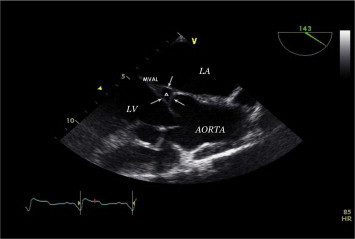
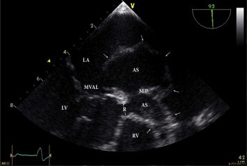
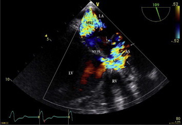
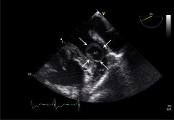
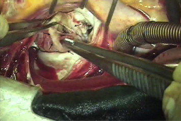
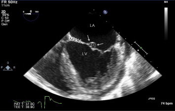
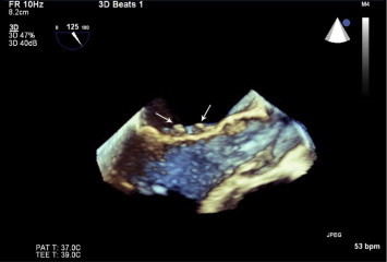
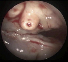
Aneurysm of the mitral valve (AMV) is rarely reported. The etiology of this unusual pathology is commonly attributed to aortic valve endocarditis (AVE) with aortic regurgitation (AR) or connective tissue disease. We present two recent cases of AMV with good correlation between pre-operative trans-esophageal echocardiography (TEE), intra-operative real-time 3-dimensional echocardiography (RT-3D-Echo) and surgical findings. The importance of diligent surveillance by TEE in patients with AVE for occurrence of AMV is emphasized. The literature on this topic is briefly reviewed.
Keywords: Aneurysm; Aortic valve endocarditis; Mitral valve; Real-time 3-dimensional echocardiography; Trans-esophageal echocardiography.
Figures

References
-
- Hotchi J., Hoshiga M., Okabe T. Impressive echocardiographic images of a mitral valve aneurysm. Circulation. 2011 April;123(14):400–402. - PubMed
-
- Michelena H.I., Suri R.M., Enriquez-Sarano M. Ruptured mycotic aneurysm of the mitral valve on real-time 3-dimensional trans-thoracic echocardiography. J Am Coll Cardiol. 2010 July 6;56(2):154. - PubMed
-
- Piazza N., Marra S., Webb J. Two cases of aneurysm of the anterior mitral valve leaflet associated with trans-catheter aortic valve endocarditis, a mere coincidence? J Thorac Cardiovasc Surg. 2010 Sept.;140(3):36–38. - PubMed
-
- Hong S.N., Perk G., Skolnick A. Evaluation of a posterior mitral valve leaflet aneurysm by real-time 3-dimensional trans-esophageal echocardiography. Echocardiography. 2009 Oct;26(9):1089–1091. - PubMed
-
- Delqado R., Martin Duran R., Vazquez de Prada J.A. The importance of the echocardiographic diagnosis of a mycotic aneurysm of the mitral septal leaflet in infective endocarditis of the aortic valve: the surgical implication. Rev Esp Cardiol. 1991 Dec;44(10):672–676. - PubMed
LinkOut - more resources
Full Text Sources
Other Literature Sources
Research Materials
