

Glioma mimicking a hypertensive intracerebral hemorrhage
- PMID: 24175027
- PMCID: PMC3809438
- DOI: 10.3340/jkns.2013.54.2.125
Glioma mimicking a hypertensive intracerebral hemorrhage
Abstract
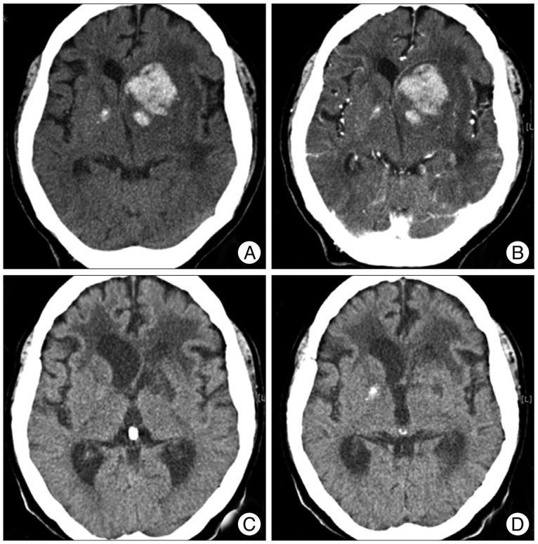
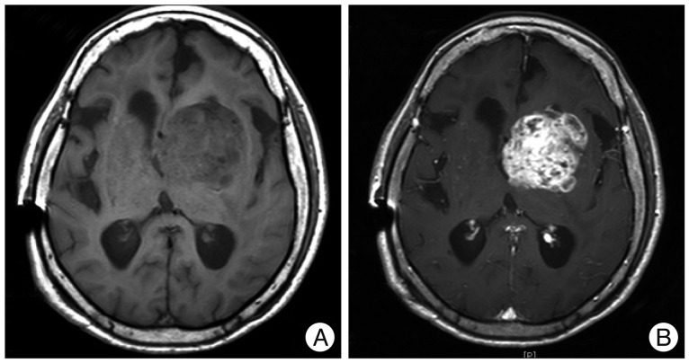
Here, we report a rare case of an anaplastic astrocytoma masquerading as a hypertensive basal ganglia hemorrhage. A 69-year-old woman who had been under medical management for hypertension during the past 3 years suddenly developed right hemiparesis with dysarthria. Brain computed tomography (CT) scans with contrast and CT angiograms revealed an intracerebral hemorrhage (ICH) in the left basal ganglia, without an underlying lesion. She was treated conservatively, but underwent a ventriculoperitoneal shunt operation 3 months after the initial attack due to deteriorated mental status and chronic hydrocephalus. Three months later, her mental status deteriorated further. Magnetic resonance imaging (MRI) with gadolinium demonstrated an irregular enhanced mass in which the previous hemorrhage occurred. The final histological diagnosis which made by stereotactic biopsy was an anaplastic astrocytoma. In the present case, the diagnosis of a high grade glioma was delayed due to tumor bleeding mimicking hypertensive ICH. Thus, a careful review of neuroradiological images including MRI with a suspicion of tumor bleeding is needed even in the patients with past medical history of hypertension.
Keywords: Anaplastic astrocytoma; Basal ganglia; Brain tumor; Hypertension; Intracerebral hemorrhage; Tumor bleeding.
Figures
Similar articles
-
Glioblastoma masquerading as a hypertensive putaminal hemorrhage: a diagnostic pitfall.Neurol Med Chir (Tokyo). 2009 Sep;49(9):427-9. doi: 10.2176/nmc.49.427. Neurol Med Chir (Tokyo). 2009. PMID: 19779291
-
MRI spot sign: Gadolinium contrast extravasation in an expanding intracerebral hematoma on MRI.Radiol Case Rep. 2019 Feb 23;14(5):535-537. doi: 10.1016/j.radcr.2019.01.018. eCollection 2019 May. Radiol Case Rep. 2019. PMID: 30976364 Free PMC article.
-
High-grade Glioma Masquerading as a Small Cerebral Hemorrhage: A Case Report.Yonago Acta Med. 2019 Oct 18;62(4):305-307. doi: 10.33160/yam.2019.11.004. eCollection 2019 Dec. Yonago Acta Med. 2019. PMID: 31849570 Free PMC article.
-
[A multicentric glioma presenting different pathological appearances: a case report].No Shinkei Geka. 2004 May;32(5):501-6. No Shinkei Geka. 2004. PMID: 15287489 Review. Japanese.
-
[A case of cystic optic glioma involving chiasma and bilateral posterior optic pathway].No Shinkei Geka. 1992 Nov;20(11):1199-204. No Shinkei Geka. 1992. PMID: 1448196 Review. Japanese.
Cited by
-
Diffuse leptomeningeal gliomatosis initially presenting with intraventricular hemorrhage: a case report and literature review.BMC Neurol. 2015 May 10;15:77. doi: 10.1186/s12883-015-0341-1. BMC Neurol. 2015. PMID: 25957575 Free PMC article. Review.
-
Revisiting prognostic factors of gliomatosis cerebri in adult-type diffuse gliomas.J Neurooncol. 2024 Jun;168(2):239-247. doi: 10.1007/s11060-024-04656-9. Epub 2024 May 3. J Neurooncol. 2024. PMID: 38700610
-
Detecting Tumor-Associated Intracranial Hemorrhage Using Proton Magnetic Resonance Spectroscopy.Neurol Int. 2024 Dec 17;16(6):1856-1877. doi: 10.3390/neurolint16060133. Neurol Int. 2024. PMID: 39728759 Free PMC article. Review.
-
Venous ectasia preceding intra-tumoral hemorrhage in a case of gliosarcoma with transverse sinus involvement.J Surg Case Rep. 2023 Jul 29;2023(7):rjad429. doi: 10.1093/jscr/rjad429. eCollection 2023 Jul. J Surg Case Rep. 2023. PMID: 37525746 Free PMC article.
-
Clinical and imaging manifestations of intracerebral hemorrhage in brain tumors and metastatic lesions: a comprehensive overview.J Neurooncol. 2024 Dec;170(3):567-578. doi: 10.1007/s11060-024-04811-2. Epub 2024 Sep 2. J Neurooncol. 2024. PMID: 39222188 Free PMC article.
References
-
- Can SM, Aydin Y, Turkmenoglu O, Aydin F, Ziyal I. Giant cell glioblastoma manifesting as traumatic intracerebral hemorrhage--case report. Neurol Med Chir (Tokyo) 2002;42:568–571. - PubMed
-
- Cemil B, Tun K, Polat O, Ozen O, Kaptanoglu E. Glioblastoma multiforme mimicking arteriovenous malformation. Turk Neurosurg. 2009;19:433–436. - PubMed
-
- Inamasu J, Kuramae T, Nakatsukasa M. Glioblastoma masquerading as a hypertensive putaminal hemorrhage : a diagnostic pitfall. Neurol Med Chir (Tokyo) 2009;49:427–429. - PubMed
-
- Kondziolka D, Bernstein M, Resch L, Tator CH, Fleming JF, Vanderlinden RG, et al. Significance of hemorrhage into brain tumors : clinicopathological study. J Neurosurg. 1987;67:852–857. - PubMed
-
- Morgenstern LB, Frankowski RF. Brain tumor masquerading as stroke. J Neurooncol. 1999;44:47–52. - PubMed
Publication types
LinkOut - more resources
Full Text Sources
Other Literature Sources
Miscellaneous
