

A complication following tooth extraction: chronic suppurative osteomyelitis
- PMID: 24175053
- PMCID: PMC3808937
A complication following tooth extraction: chronic suppurative osteomyelitis
Abstract
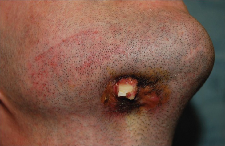
A complication following tooth extraction: a case report of chronic suppurative osteomyelitis.
Objective: This article presents a case report about the surgery treatment of chronic suppurative osteomyelitis following a tooth extraction.
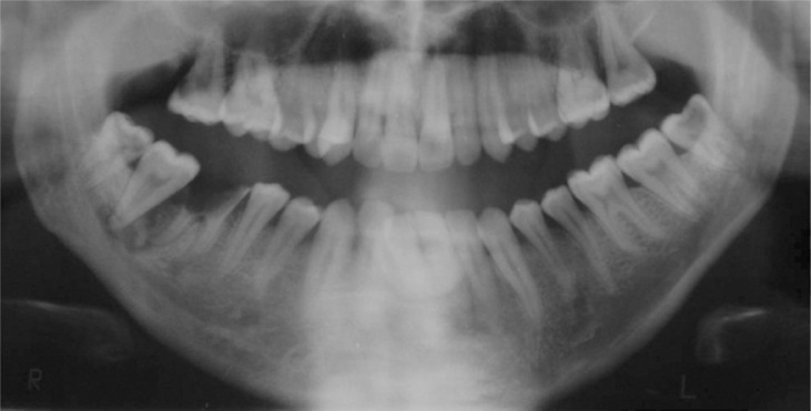
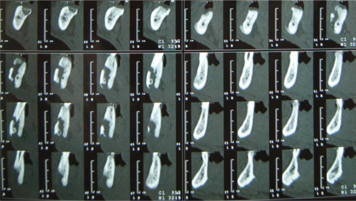
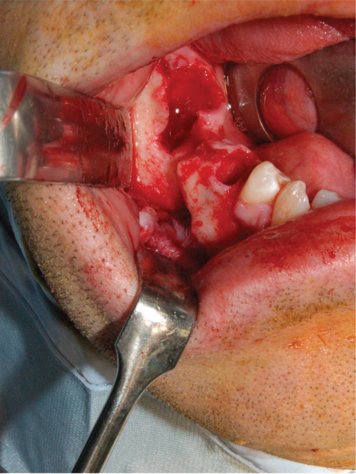
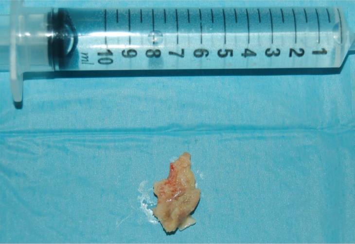
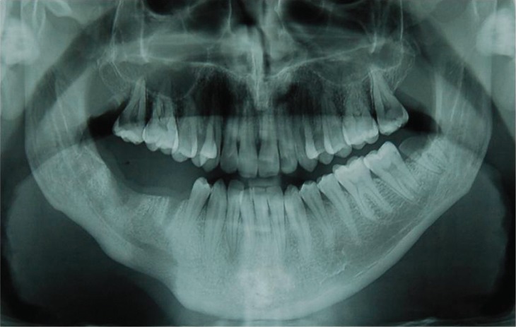
Methods: Cone beam computed tomography revealed a sequestrum bone formation that required the sequestrectomy and the debridement of the involved area. The prescription of oral penicillin and metronidazole were necessary after and before the surgery. Also 20 sessions of hyperbaric oxygen therapy were important for the healing of the marrow space.
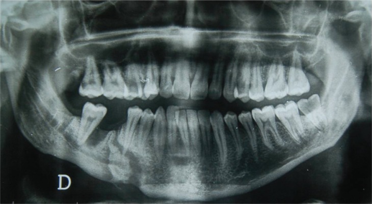
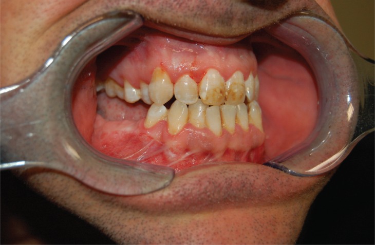
Results: The histologic test confirmed the diagnosis of "Chronic suppurative osteomyelitis". Clinically the post-operative course showed no complications but a good healing of the bone tissue. Culture reports revealed two microorganisms, streptococcus viridans and staphylococcus, that are sensitive to penicillin.
Conclusions: Clinical results confirmed the validity of the sequestrectomy and the debridement of the involved area for the treatment of chronic suppurative osteomyelitis. Such approach has always to be preferred because it guarantees the healing of bone tissue.
Keywords: bone sequestrum; chronic osteomyelitis; complication tooth extraction; sequestrectomy.
Figures

Similar articles
-
Treating low- and medium-potency bisphosphonate-related osteonecrosis of the jaws with a protocol for the treatment of chronic suppurative osteomyelitis: report of 7 cases.Oral Surg Oral Med Oral Pathol Oral Radiol Endod. 2009 Feb;107(2):e1-7. doi: 10.1016/j.tripleo.2008.09.021. Epub 2008 Dec 13. Oral Surg Oral Med Oral Pathol Oral Radiol Endod. 2009. PMID: 19071040
-
Exfoliation and simultaneous formation of condylar process following chronic osteomyelitis of the mandible.J Craniofac Surg. 2012 Jul;23(4):e319-22. doi: 10.1097/SCS.0b013e31825433f1. J Craniofac Surg. 2012. PMID: 22801167
-
Chronic suppurative osteomyelitis of mandible: a case report.Craniomaxillofac Trauma Reconstr. 2013 Sep;6(3):197-200. doi: 10.1055/s-0033-1343781. Epub 2013 May 31. Craniomaxillofac Trauma Reconstr. 2013. PMID: 24436759 Free PMC article.
-
Osteomyelitis of the jaws: a 50-year perspective.J Oral Maxillofac Surg. 1993 Dec;51(12):1294-301. doi: 10.1016/s0278-2391(10)80131-4. J Oral Maxillofac Surg. 1993. PMID: 8229407 Review.
-
[Bone disease with pain. Osteomyelitis in the long bone].Clin Calcium. 2008 Jun;18(6):836-43. Clin Calcium. 2008. PMID: 18515955 Review. Japanese.
Cited by
-
Effect of Intraoral Drainage after Impacted Mandibular Third Molar Extraction on Non-Infectious Postoperative Complications.J Clin Med. 2021 Oct 14;10(20):4705. doi: 10.3390/jcm10204705. J Clin Med. 2021. PMID: 34682828 Free PMC article.
-
A novel computationally engineered collagenase reduces the force required for tooth extraction in an ex-situ porcine jaw model.J Biol Eng. 2023 Jul 17;17(1):47. doi: 10.1186/s13036-023-00366-4. J Biol Eng. 2023. PMID: 37461028 Free PMC article.
-
Pre- and postoperative management techniques. Before and after. Part 2: the removal of third molars.Br Dent J. 2015 Mar 13;218(5):279-84. doi: 10.1038/sj.bdj.2015.145. Br Dent J. 2015. PMID: 25766164 Review.
-
Diffuse B-Cell Lymphoma of the Mandible Disguised as Acute Osteomyelitis.Eur J Case Rep Intern Med. 2024 Jan 22;11(2):004243. doi: 10.12890/2024_004243. eCollection 2024. Eur J Case Rep Intern Med. 2024. PMID: 38352811 Free PMC article.
-
Osteomyelitis of the Mandible Caused by Late Fracture following Third Molar Extraction.Case Rep Dent. 2019 Aug 4;2019:5421706. doi: 10.1155/2019/5421706. eCollection 2019. Case Rep Dent. 2019. PMID: 31467733 Free PMC article.
References
-
- Topazian RG. Osteomyelitis of jaws. In: Topazian RG, Goldberg MH, editors. Oral and maxillofacial infections. 3rd ed. Philadelphia, PA: Saunders; 1994. pp. 251–286.
-
- Yeoh SC, Mac Mahon S, Schifter M. Chronic suppurative osteomyelitis of the mandible: Case report. Aust Dent J. 2005;50:200–3. - PubMed
-
- Marx R E. Chronic osteomyelitis of the jaws. Oral Maxillofac Surg Clin North Am. 1991;3:367.
-
- Mercuri LG. Acute osteomyelitis of the jaws. Oral Maxillofac Surg Clin North Am. 1991;3:355.
-
- Baltensperger M, Eyrich G. Osteomyelitis of the Jaws: definitions and classification. In: Baltensperger M, Eyrich G, editors. Osteomyelitis of jaws. Berlin: Springer; 2009. pp. 5–56.
Publication types
LinkOut - more resources
Full Text Sources