A giant popliteal artery aneurysm treated with exclusion and bypass using a saphenous vein
- PMID: 24175274
- PMCID: PMC3810561
- DOI: 10.5090/kjtcs.2013.46.5.369
A giant popliteal artery aneurysm treated with exclusion and bypass using a saphenous vein
Abstract
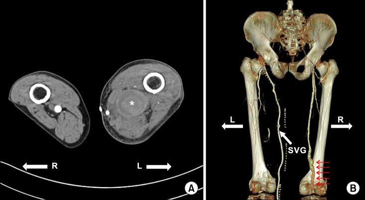
While popliteal artery aneurysm is the most common form of peripheral artery aneurysm, it is a rare condition in the general population. Furthermore, a giant popliteal artery aneurysm has not previously been reported in Korea. A 67-year-old male presented with left thigh pain that had begun 4 months earlier and was aggravated when in a sitting position. We found a giant aneurysm on the left popliteal artery and performed a bypass from the common femoral artery to the distal popliteal artery below the knee, using the autologous greater saphenous vein, and excluded the aneurysm at the sites of anastomoses.
Keywords: Endovascular procedures; Peripheral vascular disease; Popliteal artery.
Conflict of interest statement
No potential conflict of interest relevant to this article was reported.
Figures

Similar articles
-
Surgery for giant popliteal artery aneurysm with a modified Sims' position.J Vasc Surg. 1998 Feb;27(2):371-3. doi: 10.1016/s0741-5214(98)70370-2. J Vasc Surg. 1998. PMID: 9510294
-
Extended posterior approach for huge popliteal aneurysm extended to superficial femoral artery.SAGE Open Med Case Rep. 2018 Jan 9;6:2050313X17752770. doi: 10.1177/2050313X17752770. eCollection 2018. SAGE Open Med Case Rep. 2018. PMID: 29348915 Free PMC article.
-
Hybrid approach for treatment of behind the knee popliteal artery aneurysms.Vascular. 2009 Sep-Oct;17(5):290-2. doi: 10.2310/6670.2009.00019. Vascular. 2009. PMID: 19769811
-
Popliteal artery aneurysms. Factors associated with thromboembolism and graft failure.Int Angiol. 2004 Mar;23(1):54-65. Int Angiol. 2004. PMID: 15156131 Review.
-
Mycotic popliteal aneurysm rupture secondary to Campylobacter fetus.Ann Vasc Surg. 2015 Jan;29(1):122.e9-11. doi: 10.1016/j.avsg.2014.05.021. Epub 2014 Jun 12. Ann Vasc Surg. 2015. PMID: 24930978 Review.
Cited by
-
Cystic Adventitial Disease of Popliteal Artery: Case Report.Int J Angiol. 2016 Mar;25(1):68-9. doi: 10.1055/s-0034-1382810. Epub 2016 Jan 12. Int J Angiol. 2016. PMID: 26900314 Free PMC article.
References
-
- Huang Y, Gloviczki P, Noel AA, et al. Early complications and long-term outcome after open surgical treatment of popliteal artery aneurysms: is exclusion with saphenous vein bypass still the gold standard? J Vasc Surg. 2007;45:706–713. - PubMed
-
- Zimmermann A, Schoenberger T, Saeckl J, et al. Eligibility for endovascular technique and results of the surgical approach to popliteal artery aneurysms at a single center. Ann Vasc Surg. 2010;24:342–348. - PubMed
-
- Pontón A, García I, Arnáiz E, et al. Endovascular repair of a ruptured giant popliteal artery aneurysm. Ann Vasc Surg. 2009;23:412.e1–412.e4. - PubMed
-
- Gao X, Qi L, Chen B, Yu H, Li J, Zhang J. A rare case of giant popliteal artery aneurysm in a young adult. Vascular. 2011;19:342–345. - PubMed
-
- Park CB, Yoo DG, Kim CW. Bilateral popliteal artery entrapment syndrome. Korean J Thorac Cardiovasc Surg. 2007;40:136–139.
Publication types
LinkOut - more resources
Full Text Sources
Other Literature Sources