

Quadriceps and hamstrings morphology is related to walking mechanics and knee cartilage MRI relaxation times in young adults
- PMID: 24175607
- PMCID: PMC4476495
- DOI: 10.2519/jospt.2013.4486
Quadriceps and hamstrings morphology is related to walking mechanics and knee cartilage MRI relaxation times in young adults
Abstract
Study design: Controlled laboratory study using a cross-sectional design.
Objectives: To analyze the relationship of quadriceps-hamstrings and medial-lateral quadriceps anatomical cross-sectional area (ACSA) ratios with knee loads during walking and articular and meniscal cartilage composition in young, healthy subjects.
Background: Muscle forces affect knee loading during walking, but it is not known if muscle morphology is associated with walking mechanics and cartilage composition in young subjects.
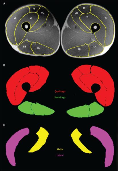
Methods: Forty-two knees from 27 young, healthy, active volunteers (age, 20-35 years; body mass index, <28 kg/m(2)) underwent 3-T magnetic resonance imaging (MRI) and 3-D motion capture. Standard MRI sequences were used for articular and meniscal cartilage T1rho and T2 relaxation times and for quadriceps and hamstrings muscle ACSA. Frontal plane kinetics during the stance phase of walking was calculated. Generalized estimating equation models were used to identify muscle variables that predicted MRI and gait parameters.
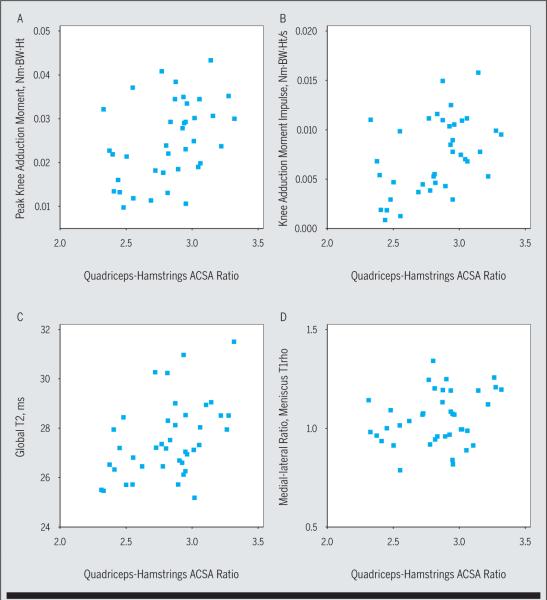
Results: Quadriceps-hamstrings and medial-lateral quadriceps ACSA ratios were positively related to frontal plane loading (β = .21-.54, P≤.006), global articular cartilage relaxation times (β = .22-.28, P≤.041), and the medial-lateral ratio of meniscus T1rho relaxation time (β = .26-.36, P≤.049). The medial-lateral quadriceps ACSA ratio was positively related to global meniscus T1rho relaxation times (β = .30, P = .046).
Conclusion: Higher quadriceps-hamstrings and medial-lateral quadriceps ACSA ratios were associated with higher frontal plane loading during walking and with articular and meniscal cartilage T1rho and T2 relaxation times. These findings highlight the relationships between different knee tissues and knee mechanics in young, healthy individuals.
Figures
References
-
- Aaboe J, Bliddal H, Alkjaer T, Boesen M, Henriksen M. The influence of radiographic severity on the relationship between muscle strength and joint loading in obese knee osteoarthritis patients. Arthritis. 2011;2011:571519. http://dx.doi.org/10.1155/2011/571519. - DOI - PMC - PubMed
-
- Akella SV, Regatte RR, Gougoutas AJ, et al. Proteoglycan-induced changes in T1rho-relaxation of articular cartilage at 4T. Magn Reson Med. 2001;46:419–423. http://dx.doi.org/10.1002/mrm.1208. - DOI - PubMed
-
- Alizai H, Nardo L, Karampinos DC, et al. Comparison of clinical semi-quantitative assessment of muscle fat infiltration with quantitative assessment using chemical shift-based water/fat separation in MR studies of the calf of post-menopausal women. Eur Radiol. 2012;22:1592–1600. http://dx.doi.org/10.1007/s00330-012-2404-7. - DOI - PMC - PubMed
-
- Andriacchi TP, Mündermann A. The role of ambulatory mechanics in the initiation and progression of knee osteoarthritis. Curr Opin Rheumatol. 2006;18:514–518. http://dx.doi.org/10.1097/01.bor.0000240365.16842.4e. - DOI - PubMed
-
- Bamman MM, Newcomer BR, Larson-Meyer DE, Weinsier RL, Hunter GR. Evaluation of the strength-size relationship in vivo using various muscle size indices. Med Sci Sports Exerc. 2000;32:1307–1313. - PubMed
Publication types
MeSH terms
Grants and funding
LinkOut - more resources
Full Text Sources
Other Literature Sources