

Periosteal thickness and cellularity in mid-diaphyseal cross-sections from human femora and tibiae of aged donors
- PMID: 24175932
- PMCID: PMC3969058
- DOI: 10.1111/joa.12133
Periosteal thickness and cellularity in mid-diaphyseal cross-sections from human femora and tibiae of aged donors
Abstract
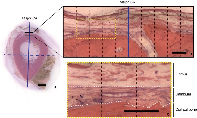
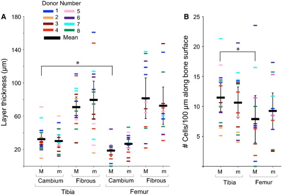
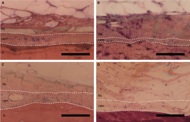
Due to lack of access in healthy patients, the structural properties underlying the inherent regenerative power and advanced material properties of the human periosteum are not well understood. Periosteum comprises a cellular cambium layer directly apposing the outer surface of bone and an outer fibrous layer encompassed by the surrounding soft tissues. As a first step to elucidating the structural and cellular characteristics of periosteum in human bone, the current study aims to measure cambium and fibrous layer thickness as well as cambium cellularity in human femora and tibiae of aged donors. The major and minor centroidal axes (CA) serve as automated reference points in cross-sections of cadaveric mid-diaphyseal femora and tibiae. Based on the results of this study, within a given individual, the cambium layer of the major CA of the tibia is significantly thicker and more cellular than the respective layer of the femur. These significant intraindividual differences do not translate to significant interindividual differences. Further, mid-diaphyseal periosteal measures including cambium and fibrous layer thickness and cellularity do not correlate significantly with age or body mass. Finally, qualitative observations of periosteum in amputated and contralateral or proximal long bones of the lower extremity show stark changes in layer organization, thickness, and cellularity. In a translational context, these novel data, though inherently limited by availability and accessibility of human mid-diaphyseal periosteum tissue, provide important reference values for the use of periosteum in the context of facilitated healing and regeneration of tissue.
Keywords: amputee; loading history; mechanobiology; periosteal cells; periosteum.
© 2013 Anatomical Society.
Figures
References
-
- Allen MR, Hock JM, Burr DB. Periosteum: biology, regulation, and response to osteoporosis therapies. Bone. 2004;35:1003–1012. doi: 10.1016/j.bone.2004.07.014. - DOI - PubMed
-
- Bertram JE, Polevoy Y, Cullinane DM. Mechanics of avian fibrous periosteum: tensile and adhesion properties during growth. Bone. 1998;22:669–675. - PubMed
-
- Carpenter RD, Carter DR. The mechanobiological effects of periosteal surface loads. Biomech Model Mechanobiol. 2008;7:227–242. - PubMed
-
- Chang H, Knothe Tate ML. Concise review: the periosteum: tapping into a reservoir of clinically useful progenitor cells. Stem Cells Transl Med. 2012;1:480–491. doi: 10.5966/sctm.2011-0056. - DOI - PMC - PubMed
-
- Chang H, Docheva D, Knothe UR, et al. 2013. Arthritic periosteal tissue from joint replacement surgery as an autologous source of stem cells In Review.
MeSH terms
LinkOut - more resources
Full Text Sources
Other Literature Sources
Medical
Research Materials
