. 2013 Nov 1;4(1):19.
doi: 10.1186/2042-6410-4-19.

Sex differences in the expression of lupus-associated miRNAs in splenocytes from lupus-prone NZB/WF1 mice

Affiliations

Sex differences in the expression of lupus-associated miRNAs in splenocytes from lupus-prone NZB/WF1 mice

Rujuan Dai et al. Biol Sex Differ. .

Abstract

Background: A majority of autoimmune diseases, including systemic lupus erythematosus (SLE), occur predominantly in females. Recent studies have identified specific dysregulated microRNAs (miRNAs) in both human and murine lupus, implying an important contribution of these miRNAs to lupus pathogenesis. However, to date, there is no study that examined sex differences in miRNA expression in immune cells as a plausible basis for sex differences in autoimmune disease. This study addresses this aspect in NZB/WF1 mice, a classical murine lupus model with marked female bias, and further investigates estrogen regulation of lupus-associated miRNAs.

Methods: The Taqman miRNA assay system was used to quantify the miRNA expression in splenocytes from male and female NZB/WF1 mice at 17-18, 23, and 30 weeks (wks) of age. To evaluate potential estrogen's effect on lupus-associated miRNAs, 6-wk-old NZB/WF1 male mice were orchidectomized and surgically implanted with empty (placebo) or estrogen implants for 4 and 26 wks, respectively. To assess the lupus status in the NZB/WF1 mice, serum anti-dsDNA autoantibody levels, proteinuria, and renal histological changes were determined.

Results: The sex differences in the expression of lupus-associated miRNAs, including the miR-182-96-183 cluster, miR-155, miR-31, miR-148a, miR-127, and miR-379, were markedly evident after the onset of lupus, especially at 30 wks of age when female NZB/WF1 mice manifested moderate to severe lupus when compared to their male counterparts. Our limited data also suggested that estrogen treatment increased the expression of aforementioned lupus-associated miRNAs, with the exception of miR-155, in orchidectomized male NZB/WF1 mice to a similar level in age-matched intact female NZB/WF1 mice. It is noteworthy that orchiectomy, itself, did not affect the expression of lupus-associated miRNAs.

Conclusion: To our knowledge, this is the first study that demonstrated sex differences in the expression of lupus-associated miRNAs in splenocytes, especially in the context of autoimmunity. The increased expression of lupus-associated miRNA in female NZB/WF1 mice and conceivably in estrogen-treated orchidectomized male NZB/WF1 mice was associated with lupus manifestation. The notable increase of lupus-associated miRNAs in diseased, female NZB/WF1 mice may be a result of both lupus manifestation and the female gender.


Figures

Figure 1
Figure 1
Comparable lupus-associated miRNA levels in splenocytes from male and female NZB/WF1 mice before lupus onset. (A) Real-time RT-PCR analysis of select lupus-associated miRNAs. The graph represents the means ± SEMs (n = 4 each). Student t test was performed (male vs female). *p < 0.05. (B) ELISA assay of serum anti-dsDNA autoantibodies. The serum samples from three non-autoimmune C57/BL6 mice were included as negative control. The mean serum anti-DNA autoantibody value in each group was indicated by black line.
Figure 2
Figure 2
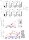
Sex differences in the expression of lupus-associated miRNAs become evident after onset of lupus. (A) Real-time RT-PCR analysis of lupus-associated miRNAs expression in splenocytes. The expression level of a specific miRNA in 23-wk-old female and 30-wk-old male and female NZB/WF1 mice was shown as the relative expression level to 23-wk-old male mice. The graphs represent the means ± SEMs (n = 5 each for 23-wk-old male and female mice groups, and n = 4 each for 30-wk-old male and female mice groups). Student t test was performed (age-matched male vs female, and sex-matched 23 wks vs 30 wks). *p < 0.05 and **p <0.01. (B and C) Anti-dsDNA autoantibody ELISA. Anti-dsDNA antibody levels were monitored sequentially every 2–4 wks in individual male (dashed blue hue lines) and female (solid pink hue line) NZB/WF1 mice that were terminated at (B) 23 wks of age and (C) 30 wks of age, respectively.
Figure 3
Figure 3
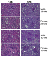
Renal pathology of male and female NZB/WF1 mice. Renal sections from 23-wk-old and 30-wk-old male and female NZB/WF1 mice were stained with H&E and PAS, respectively. Representative images from each age and gender group were shown. Note that only the 30-wk-old female NZB/WF1 mice demonstrated moderate to severe mesangioproliferative glomerulonephritis and tubular protein casts (indicated by arrow). The mice in the other groups showed no or minimal glomerular inflammation.
Figure 4
Figure 4
Estrogen regulation of lupus-associated miRNAs expression in splenocytes from orchidectomized NZB/WF1 mice. (A) Real-time RT-PCR analysis of miRNA expression in splenocytes from 10-wk-old and 32-wk-old intact male, placebo- and estrogen-treated orchidectomized male NZB/WF1 mice. The expression level of a specific miRNA in all other groups was shown as relative level to the 10-wk-old intact male mice. The graphs represent the means ± SEMs (n = 4 each for 10-wk-old intact and placebo-treated mice groups, n = 5 each for 10-wk-old estrogen-treated, 32-wk-old intact, and 32-wk-old placebo-treated mice groups, and n = 2 for 32-wk-old estrogen-treated mice group). One-way ANOVA with Tukey-Kramer all pair's comparison tests were performed to compare the expression among the age-matched intact, placebo-, and estrogen-treated mice. Asterisk indicates the statistical significance between the age-matched placebo control and estrogen-treated mice, and the number sign indicates the statistical significance between the age-matched intact control and estrogen-treated mice. Asterisk and number sign depict p < 0.05; double asterisk and double number sign depict p < 0.01. (B) Anti-dsDNA autoantibody ELISA. The figure shows the serum anti-DNA autoantibody levels in NZB/WF1 male mice from aforementioned six treatment groups. Four C57BL6/ mice were included as negative controls. The bold black line represents the mean serum autoantibody value of each group.
Figure 5
Figure 5
Estrogen increased the expression of miR-223 and miR-451 expression in splenocytes from NZB/WF1 mice. The graphs show increased miR-223 and miR-451 expression in splenocytes from 10-wk-old (A) and 32-wk-old (B) estrogen-treated orchidectomized NZB/WF1 mice when compared to either age-matched placebo control or intact control mice. The graph shows the means ± SEMS (n = 2 for 32-wk-old, estrogen-treated mice group, and n = 4 each for the other groups). Student t test was performed (estrogen vs placebo or intact). *p < 0.05 and *** p <0.001.
Figure 6
Figure 6
Comparable lupus-associated miRNAs levels in splenocytes from estrogen-treated male and age-matched intact female NZB/WF1 mice. The expression levels of miRNA in 30-wk-old intact female and 32-wk-old estrogen-treated orchidectomized NZB/WF1 mice were shown as the relative expression levels to 30-wk-old intact male mice. The graph shows the means ± SEMS (n = 2 for 32-wk-old estrogen-treated mice group, and n = 4 each for 30-wk-old intact male and female mice groups). Student t test was performed (30-wk-old female NZB/WF1 vs 32-wk-old estrogen-treated orchidectomized NZB/WF1). *p < 0.05.

References

    1. Wijchers PJ, Festenstein RJ. Epigenetic regulation of autosomal gene expression by sex chromosomes. Trends Genet. 2011;27:132–140. doi: 10.1016/j.tig.2011.01.004. - DOI - PubMed
    1. Libert C, Dejager L, Pinheiro I. The X chromosome in immune functions: when a chromosome makes the difference. Nat Rev Immunol. 2010;10:594–604. doi: 10.1038/nri2815. - DOI - PubMed
    1. Arnold AP, Lusis AJ. Understanding the sexome: measuring and reporting sex differences in gene systems. Endocrinology. 2012;153:2551–2555. doi: 10.1210/en.2011-2134. - DOI - PMC - PubMed
    1. Morgan CP, Bale TL. Sex differences in microRNA regulation of gene expression: no smoke, just miRs. Biol Sex Differ. 2012;3:22. doi: 10.1186/2042-6410-3-22. - DOI - PMC - PubMed
    1. Hedrich CM, Tsokos GC. Epigenetic mechanisms in systemic lupus erythematosus and other autoimmune diseases. Trends Mol Med. 2011;17:714–724. doi: 10.1016/j.molmed.2011.07.005. - DOI - PMC - PubMed

LinkOut - more resources