. 2014 Jan;91(1):26-38.
doi: 10.1111/mmi.12447. Epub 2013 Nov 24.

Crystal structure of an HD-GYP domain cyclic-di-GMP phosphodiesterase reveals an enzyme with a novel trinuclear catalytic iron centre

Affiliations
Free PMC article

Crystal structure of an HD-GYP domain cyclic-di-GMP phosphodiesterase reveals an enzyme with a novel trinuclear catalytic iron centre

Dom Bellini et al. Mol Microbiol. 2014 Jan.
Free PMC article

Abstract

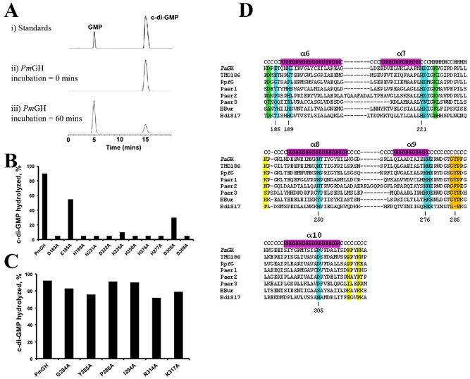
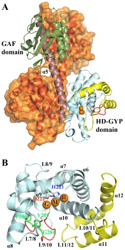
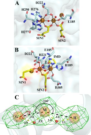
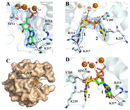
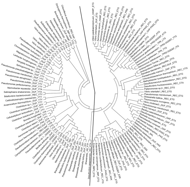
Bis-(3',5') cyclic di-guanylate (c-di-GMP) is a key bacterial second messenger that is implicated in the regulation of many crucial processes that include biofilm formation, motility and virulence. Cellular levels of c-di-GMP are controlled through synthesis by GGDEF domain diguanylate cyclases and degradation by two classes of phosphodiesterase with EAL or HD-GYP domains. Here, we have determined the structure of an enzymatically active HD-GYP domain protein from Persephonella marina (PmGH) alone, in complex with substrate (c-di-GMP) and final reaction product (GMP). The structures reveal a novel trinuclear iron binding site, which is implicated in catalysis and identify residues involved in recognition of c-di-GMP. This structure completes the picture of all domains involved in c-di-GMP metabolism and reveals that the HD-GYP family splits into two distinct subgroups containing bi- and trinuclear metal centres.


Figures

Figure 1
Figure 1
PDE activity of purified HD‐GYP domain PmGH and variants with alanine substitutions. A. Representative HPLC traces showing standards (i), aliquots of reaction mixtures boiled at 0 min (ii) and after 60 min (iii) incubation with the PmGH protein. The identity of the product was confirmed by mass spectrometry. B. Effects of alanine substitutions in metal ligands (E185, H189, H221, D222, H250, H276, H277 and D305), a strongly conserved residue in the family (D308) and the two putative catalytic residues (D183 and K225) on cyclic di‐GMP hydrolysis. C. Effects of alanine substitutions in residues in the GYP motif (G284, Y285 and P286), the conserved I294 position and other residues involved in substrate binding (R314 and K317) on cyclic di‐GMP hydrolysis. D. Primary sequence alignment of HD‐GYP domains of PmGH with some of the most well‐characterized HD‐GYP proteins, such as TM0186 (Thermotoga maritima), RpfG (Xanthomonas campestris pv. campestris), Paer1‐3 (Pseudomanas aeruginosa), BBur (Borrelia burgdorferi) and Bd1817 (Bdellovibrio bacteriovorus). Metal ligands, catalytic residues, substrate ligands and GYP motif, based on the PmGH structure, are highlighted in cyan, green, yellow and orange respectively.
Figure 2
Figure 2
Structure of PmGH. A. Structure of the PmGH homodimer. Molecule A of the dimer is shown in ribbon representation with the GAF domain in green, the long inter‐domain dimerization helix α5 in maroon, the core HD domain in cyan, the GYP motif‐containing loop in red and the additional surface decorating α‐ helices which complete the HD‐GYP domain in yellow. Molecule B of the dimer is shown in ribbon representation in orange with a semi‐transparent surface. The trinuclear iron centre is shown as orange spheres. B. Detailed view of the HD‐GYP domain of PmGH in ribbon presentation. Colour codes as in (A) with the addition of the HD and GYP motif residues shown in ball and stick. Labelling for α‐helices and turns are shown. The central metal iron has been labelled as the middle site (M) and the two flanking metal sites as H and G, to reflect their proximity to the HD and GYP motifs respectively.
Figure 3
Figure 3
The tri‐iron metal centre of PmGH. Detailed views of the tri‐iron centre showing the first co‐ordination sphere for the GM metal pair (A) and for the MH metal pair (B) with the co‐ordination of the H and G sites not shown in (A) and (B) respectively for clarity. Protein metal interactions are highlighted as black lines. Fe atoms are shown as orange spheres. Protein side‐chain metal ligands are in stick mode, coloured by atom type with carbon in pelican, while the carbon atoms of the metal ligands from the crystallization buffer, 2 succinates SIN‐1,2 and an imidazole ion (IMD), are in yellow. (C) Fe‐specific difference DANO map (Than et al., 2005) in green and Mn anomalous difference map in red, both contoured at 0.043 eÅ‐3. The angle subtended by the tri‐iron centre is shown. Black dashed lines depict FeFe distances, while yellow dashed lines indicate bond distances for the pair of μ‐hydroxides. Bond distances are in Ångstroms.
Figure 4
Figure 4
Substrate binding by PmGH. A. View of GMP shown in stick mode and coloured by atom type bound to PmGH. Bonding interactions are represented by dashed lines with distances in Angstroms. Difference electron density map for GMP is contoured at 2 σ. B. View of cyclic di‐GMP bound to a metal depleted subunit of PmGH. Electron density for cyclic di‐GMP is from a bias‐removed omit map contoured at 2 σ. Fe atoms occupying the middle (M) and HD (H) sites are shown as semi transparent spheres as they are not present in the subunit (due to chelation by EDTA), and are taken from superposition of the equivalent metals from the high resolution structure of PmGH. C. Surface representation of the PmGH HDGYP domain monomer subunit showing the binding cavity for cyclic‐di‐GMP, which is represented in stick mode and coloured by atom type. D. Superposition of the structures of PmGH bound to cyclic di‐GMP and GMP. Both nucleotides are shown in stick mode.
Figure 5
Figure 5
Comparison of the structures of the HD‐GYP domain from PmGH and the unconventional HD‐GYP domain of Bd1817. A. PmGH and Bd1817 PDE domains are shown in ribbon representation and coloured cyan and olive green respectively. The PmGH metal centre is as for Fig. 1, while the Bd1817 binuclear Fe centre with a bridging hydroxide ion is shown as green spheres. The shift in orientation in helices α6 and α10 in PmGH when compared with Bd1817 which allows the protein to accommodate the trinuclear metal centre is highlighted. B. Comparison of the HD‐GYP domain nucleotide‐binding pocket highlighting the opened and closed conformations observed for PmGH and Bd1817 in red and green respectively. Hydrogen bonds proposed to hold the Bd1817 in this close conformation are shown as black dashed lines. The double headed black arrow depicts the distance in Angstroms between the L7/8 and L10/11 loops in PmGH for which the active site is in an open conformation. C. Secondary structure superposition using protein fragments ranging from first to last residue involved in metal co‐ordination of PmGH and Bd1817 showing only the protein metal ligands.
Figure 6
Figure 6
Maximum parsimony analysis of various HD‐GYP domain sequences. The evolutionary history was inferred using the maximum parsimony (MP) method. Tree #1 out of 17 most parsimonious trees (length = 3871) is shown. The consistency index is 0.257040 (0.255115), the retention index is 0.444680 (0.444680), and the composite index is 0.114300 (0.113445) for all sites and parsimony‐informative sites (in parentheses). The MP tree was obtained using the Close‐Neighbor‐Interchange algorithm (Suzuki et al., 2002) with search level 0 in which the initial trees were obtained with the random addition of sequences (10 replicates). The analysis involved 122 amino acid sequences. All positions containing gaps and missing data were eliminated. There were a total of 116 positions in the final dataset. Sequence labels are in the following format: (1) organism name followed by a number if more than one sequence is present from the same organism; (2) a +/− sign indicating Gram + or Gram −; (3) other domains present besides the HD‐GYP domain are indicated, followed by a number in case of multiple copies; (4) a 3 letter code for the 3 residues triplet subfamily signature corresponding to positions 185–187 in PmGH. Extra domains are: REC = CheYhomologous receiver domain; GAF = present in cyclic di‐GMP phosophodiesterase, Adenyl cyclase, Fhla; PAS = present in Periodic circadian protein, Ah receptor nuclear translocator protein, Single‐minded protein; HAMP = present in Histidine kinases, Adenyl cyclases, Methyl‐accepting proteins and Phosphatases; HD = extra HD domain of unknown function missing the GYP motif; TPR = domain containing the Teratrico Peptide Repeat region; GGDEF = diguanylate cyclase containing the GGDEF motif; SBP = bacterial extracellular Solute‐ Binding Protein; DUF = Domain of Unknown Function. Evolutionary analyses were conducted in Mega5 (Tamura et al., 2011).

Comment in

References

    1. Albeck, S., Alzari, P., Andreini, C., Banci, L., Berry, I.M., Bertini, I., et al (2006) SPINE bioinformatics and data‐management aspects of high‐throughput structural biology. Acta Crystallogr D Biol Crystallogr 62: 1184–1195 - PMC - PubMed
    1. Aravind, L., and Koonin, E.V. (1998) The HD domain defines a new superfamily of metal‐dependent phosphohydrolases. Trends Biochem Sci 23: 469–472 - PubMed
    1. Barends, T.R., Hartmann, E., Griese, J.J., Beitlich, T., Kirienko, N.V., Ryjenkov, D.A., et al (2009) Structure and mechanism of a bacterial light‐regulated cyclic nucleotide phosphodiesterase. Nature 459: 1015–1018 - PubMed
    1. Bellini, D., Fordham‐Skelton, A., and Papiz, M.Z. (2011) STRU‐cloning: a fast, inexpensive and efficient cloning procedure applicable to both small scale and structural genomics size cloning. Mol Biotechnol 48: 30–37 - PubMed
    1. Boyd, C.D., and O'Toole, G.A. (2012) Second messenger regulation of biofilm formation: breakthroughs in understanding c‐di‐GMP effector systems. Annu Rev Cell Dev Biol 28: 439–462 - PMC - PubMed

Publication types