

Neurochemical characterisation of lamina II inhibitory interneurons that express GFP in the PrP-GFP mouse
- PMID: 24176114
- PMCID: PMC4228398
- DOI: 10.1186/1744-8069-9-56
Neurochemical characterisation of lamina II inhibitory interneurons that express GFP in the PrP-GFP mouse
Abstract
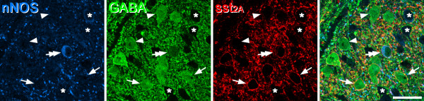
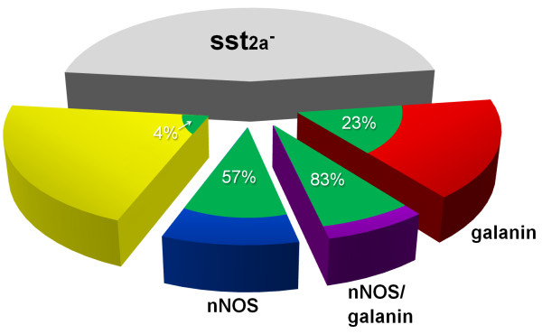
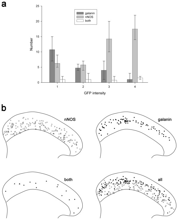
Background: Inhibitory interneurons in the superficial dorsal horn play important roles in modulating sensory transmission, and these roles are thought to be performed by distinct functional populations. We have identified 4 non-overlapping classes among the inhibitory interneurons in the rat, defined by the presence of galanin, neuropeptide Y, neuronal nitric oxide synthase (nNOS) and parvalbumin. The somatostatin receptor sst2A is expressed by ~50% of the inhibitory interneurons in this region, and is particularly associated with nNOS- and galanin-expressing cells. The main aim of the present study was to test whether a genetically-defined population of inhibitory interneurons, those expressing green fluorescent protein (GFP) in the PrP-GFP mouse, belonged to one or more of the neurochemical classes identified in the rat.
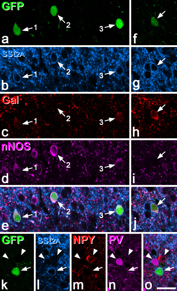
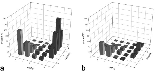
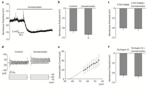
Results: The expression of sst2A and its relation to other neurochemical markers in the mouse was similar to that in the rat, except that a significant number of cells co-expressed nNOS and galanin. The PrP-GFP cells were entirely contained within the set of inhibitory interneurons that possessed sst2A receptors, and virtually all expressed nNOS and/or galanin. GFP was present in ~3-4% of neurons in the superficial dorsal horn, corresponding to ~16% of the inhibitory interneurons in this region. Consistent with their sst2A-immunoreactivity, all of the GFP cells were hyperpolarised by somatostatin, and this was prevented by administration of a selective sst2 receptor antagonist or a blocker of G-protein-coupled inwardly rectifying K+ channels.
Conclusions: These findings support the view that neurochemistry provides a valuable way of classifying inhibitory interneurons in the superficial laminae. Together with previous evidence that the PrP-GFP cells form a relatively homogeneous population in terms of their physiological properties, they suggest that these neurons have specific roles in processing sensory information in the dorsal horn.
Figures
References
-
- Polgár E, Hughes DI, Riddell JS, Maxwell DJ, Puskar Z, Todd AJ. Selective loss of spinal GABAergic or glycinergic neurons is not necessary for development of thermal hyperalgesia in the chronic constriction injury model of neuropathic pain. Pain. 2003;104:229–239. doi: 10.1016/S0304-3959(03)00011-3. - DOI - PubMed
Publication types
MeSH terms
Substances
Grants and funding
LinkOut - more resources
Full Text Sources
Other Literature Sources
Research Materials
