Intrinsic foot muscle deterioration is associated with metatarsophalangeal joint angle in people with diabetes and neuropathy
- PMID: 24176198
- PMCID: PMC3893062
- DOI: 10.1016/j.clinbiomech.2013.10.006
Intrinsic foot muscle deterioration is associated with metatarsophalangeal joint angle in people with diabetes and neuropathy
Abstract
Background: Metatarsophalangeal joint deformity is associated with skin breakdown and amputation. The aims of this study were to compare intrinsic foot muscle deterioration ratios (ratio of adipose to muscle volume), and physical performance in subjects with diabetic neuropathy to controls, and determine their associations with 1) metatarsophalangeal joint angle and 2) history of foot ulcer.
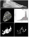
Methods: 23 diabetic, neuropathic subjects [59 (SD 10) years] and 12 age-matched controls [57 (SD 14) years] were studied. Radiographs and MRI were used to measure metatarsophalangeal joint angle and intrinsic foot muscle deterioration through tissue segmentation by image signal intensity. The Foot and Ankle Ability Measure evaluated physical performance.
Findings: The diabetic, neuropathic group had a higher muscle deterioration ratio [1.6 (SD 1.2) vs. 0.3 (SD 0.2), P<0.001], and lower Foot and Ankle Ability Measure scores [65.1 (SD 24.4) vs. 98.3 (SD 3.3) %, P<0.01]. The correlation between muscle deterioration ratio and metatarsophalangeal joint angle was r=-0.51 (P=0.01) for all diabetic, neuropathic subjects, but increased to r=-0.81 (P<0.01) when only subjects with muscle deterioration ratios >1.0 were included. Muscle deterioration ratios in individuals with diabetic neuropathy were higher for those with a history of ulcers.
Interpretation: Individuals with diabetic neuropathy had increased intrinsic foot muscle deterioration, which was associated with second metatarsophalangeal joint angle and history of ulceration. Additional research is required to understand how foot muscle deterioration interacts with other impairments leading to forefoot deformity and skin breakdown.
Keywords: Foot deformity; Intermuscular adipose tissue; Muscle.
© 2013.
Figures


References
-
- Ahroni JH, Boyko EJ, Forsberg RC. Clinical correlates of plantar pressure among diabetic veterans. Diabetes Care. 1999;22:965–972. - PubMed
-
- Andersen H, Gjerstad MD, Jakobsen J. Atrophy of foot muscles: a measure of diabetic neuropathy. Diabetes Care. 2004;27:2382–2385. - PubMed
-
- Andreassen CS, Jakobsen J, Ringgaard S, Ejskjaer N, Andersen H. Accelerated atrophy of lower leg and foot muscles- a follow-up study of long-term diabetic polyneuropathy using magnetic resonance imaging (MRI) Diabetologia. 2009;52:1182–1191. - PubMed
-
- Boulton AJ. The pathogenesis of diabetic foot problems: an overview. Diabet Med. 1996;13(Suppl 1):S12–16. - PubMed
Publication types
MeSH terms
Grants and funding
LinkOut - more resources
Full Text Sources
Other Literature Sources
Medical

