. 2013 Oct 31;4(10):e894.
doi: 10.1038/cddis.2013.401.

killerFLIP: a novel lytic peptide specifically inducing cancer cell death

Affiliations

killerFLIP: a novel lytic peptide specifically inducing cancer cell death

B Pennarun et al. Cell Death Dis. .

Abstract

One of the objectives in the development of effective cancer therapy is induction of tumor-selective cell death. Toward this end, we have identified a small peptide that, when introduced into cells via a TAT cell-delivery system, shows a remarkably potent cytoxicity in a variety of cancer cell lines and inhibits tumor growth in vivo, whereas sparing normal cells and tissues. This fusion peptide was named killerFLIP as its sequence was derived from the C-terminal domain of c-FLIP, an anti-apoptotic protein. Using structure activity analysis, we determined the minimal bioactive core of killerFLIP, namely killerFLIP-E. Structural analysis of cells using electron microscopy demonstrated that killerFLIP-E triggers cell death accompanied by rapid (within minutes) plasma membrane permeabilization. Studies of the structure of the active core of killerFLIP (-E) indicated that it possesses amphiphilic properties and self-assembles into micellar structures in aqueous solution. The biochemical properties of killerFLIP are comparable to those of cationic lytic peptides, which participate in defense against pathogens and have also demonstrated anticancer properties. We show that the pro-cell death effects of killerFLIP are independent of its sequence similarity with c-FLIPL as killerFLIP-induced cell death was largely apoptosis and necroptosis independent. A killerFLIP-E variant containing a scrambled c-FLIPL motif indeed induced similar cell death, suggesting the importance of the c-FLIPL residues but not of their sequence. Thus, we report the discovery of a promising synthetic peptide with novel anticancer activity in vitro and in vivo.


Figures

Figure 1
Figure 1
The fusion peptide induces cell death in Jurkat cells. (a) Treatment with fusion peptide decreased cell viability as assessed by relative ATP levels. Jurkat cells were treated overnight with various concentrations of TAT or fusion peptide before ATP levels quantification (expressed as percentage relative to the control). (b) Treatment with fusion peptide-induced cell death as assessed by Trypan blue exclusion assay. Jurkat cells were treated overnight with various concentrations of TAT or fusion peptide before Trypan blue staining and quantification. Data representative of at least two independent experiments performed in triplicate were shown. Values are expressed as mean (± S.D.)
Figure 2
Figure 2
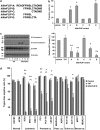
killerFLIP-E is the bioactive motif of the fusion peptide. (a) Amino-acid sequences of the five c-FLIPL-derived motifs tested. (b) Jurkat cells were treated with the killerFLIP variants (15 μM) for 18 h before quantification of cell death with PI staining and flow cytometer analysis (top panel). Values are expressed as mean (±S.D.) of three independent experiments. *P<0.001 using Student's t-test for the comparison between TAT-treated and cells treated with the killerFLIP variants. Jurkat cells were treated with the various peptides (15 μM) for 18 h before ATP quantification (bottom panel). Values are expressed as mean (± S.D.) of four independent experiments. *P<0.05 and **P<0.005 using Student's t-test for the comparison between TAT-treated and cells treated with the killerFLIP variants. (c) Western blot analysis showing time-dependent effects of TAT and killerFLIP-E (both 15 μM) on caspase 3 and PARP-1 cleavage in Jurkat cells; *Indicates an aspecific band. Data are representative of three independent experiments. (d) Effects of 18 h killerFLIP-E treatment (20 μM) on cell death as assessed by Trypan blue exclusion assay in a panel of normal and cancer cell lines; *P<0.01 and **P<0.001 (Student's t-test). Data represent mean (± S.D.) of at least two independent experiments
Figure 3
Figure 3
killerFLIP-E rapidly disrupts plasma membrane integrity and induces ultrastructural alterations in cancer cells. (a) Jurkat and (b) K562 cells were treated with 20 μM TAT or killerFLIP-E for the indicated times before Trypan blue exclusion assay. Values expressed as mean (± S.D.). (c) EM micrographs showing differences in Jurkat cell morphology upon exposure to 20 μM TAT (left) and killerFLIP-E (right) for 5 min
Figure 4
Figure 4
The hydrophobic profile of killerFLIP-E is more important than its sequence for the effects on cell death. (a) The hydrophobic profiles of TAT and killerFLIP-E were analyzed. (b) Measurement of TAT and killerFLIP-E particule size using DLS. The mean (± S.D.) is shown (n ≥7). (c) killerFLIP-E and scrambled killerFLIP-E had similar effects on cell death in Jurkat cells as assessed by relative ATP levels. Cells were treated for 18 h with TAT, killerFLIP-E or a scrambled killerFLIP-E before quantification of the relative ATP levels
Figure 5
Figure 5
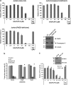
killerFLIP-E induces cell death independently of apoptosis and necroptosis. (a) Effects of TAT or killerFLIP-E in parental Jurkat cells, FADD-deficient Jurkat cells and caspase 8-deficient Jurkat cells as assessed by ATP levels. The results are expressed as the percentage of ATP signal in treated cells relative to vehicle-treated controls. Lack of caspase 8 and FADD protein expression in caspase 8-deficient (caspase 8 def.) and FADD-deficient (FADD def.) Jurkat cells was confirmed by western blot analysis; *Indicates an aspecific band. (b) Effects of pre-treatment with various concentrations of the pancaspase inhibitor zVAD-fmk on cell death induced by vehicle, TAT (10 μM), killerFLIP-E (7.5 μM) or HGS-ETR2 (1 μg/ml) in Jurkat cells, as assessed by Trypan blue staining and quantification. Cells were preincubated with zVAD-fmk for 30 min before 17 h treatment with TAT, killerFLIP-E or HGS-ETR2. Data representative of at least three independent experiments. *P<0.001 using Student's t-test for the comparison between vehicle-treated groups and cells pre-incubated with zVAD-fmk. (c) Parental Jurkat cells (SVT35) and RIPK1 −/− cells were treated with various concentrations of killerFLIP-E or 15 μM TAT before relative ATP level quantification. Data representative of two independent experiments are shown as mean (± S.D.). Lack of RIPK1 protein expression in RIPK1 deficient (RIPK1 def.) Jurkat cells was confirmed by western blot analysis
Figure 6
Figure 6
killerFLIP was well tolerated in mice. (a) Box plots showing weights of experimental animals before and after treatment with the indicated peptides. Four groups of eight mice each were weighed on the first and last days (day 18) of treatment. Black dots represent mean for each treatment group. (b) Representative liver sections from the indicated experimental groups of mice stained with H&E
Figure 7
Figure 7
Growth of established HCT116 xenograft is significantly impaired in response to killerFLIP. (a) Mice subcutaneously implanted with HCT116 xenograft were intraperitoneally administered once per day with the indicated doses of TAT or killerFLIP for 18 days. Tumor size was measured as described in Material and Methods section. (b) Treatment with killerFLIP-induced cell death in xenograft tumors. Histological analysis of biopsy samples from the mice treated with vehicle, TAT or killerFLIP. Tumor sections from untreated or mice injected with TAT or killerFLIP for 18 days were stained using TUNEL assay

References

    1. Alderton GK, Bordon Y. Tumour immunotherapy--leukocytes take up the fight. Nat Rev Immunol. 2012;12:237. - PubMed
    1. Pecot CV, Calin GA, Coleman RL, Lopez-Berestein G, Sood AK. RNA interference in the clinic: challenges and future directions. Nat Rev Cancer. 2011;11:59–67. - PMC - PubMed
    1. Vander Heiden MG. Targeting cancer metabolism: a therapeutic window opens. Nat Rev Drug Discov. 2011;10:671–684. - PubMed
    1. Tu Z, Hao J, Kharidia R, Meng XG, Liang JF. Improved stability and selectivity of lytic peptides through self-assembly. Biochem Biophys Res Commun. 2007;361:712–717. - PubMed
    1. Papo N, Shai Y. New lytic peptides based on the D,L-amphipathic helix motif preferentially kill tumor cells compared to normal cells. Biochemistry. 2003;42:9346–9354. - PubMed

Publication types

LinkOut - more resources