

Evaluation of swallow function after tongue cancer treatment using real-time magnetic resonance imaging: a pilot study
- PMID: 24177574
- PMCID: PMC5110428
- DOI: 10.1001/jamaoto.2013.5444
Evaluation of swallow function after tongue cancer treatment using real-time magnetic resonance imaging: a pilot study
Abstract
Importance: Magnetic resonance imaging (MRI) has the advantage of imaging swallow function at any anatomical level without changing the position of patient, which can provide detailed information than modified barium swallow, by far the gold standard of swallow evaluation.
Objective: To investigate the use of real-time MRI in the evaluation of swallow function of patients with tongue cancer.
Design, setting, and participants: Real-time MRI experiments were performed on a Signa Excite HD 1.5-T scanner (GE Healthcare), with gradients capable of 40-mT/m (milli-Tesla per meter) amplitudes and 150-mT/m/ms (mT/m per millisecond) slew rates. The sequence used was spiral fast gradient echo sequence. Four men with base of tongue or oral tongue squamous cell carcinoma and 3 age-matched healthy men with normal swallowing participated in the experiment.
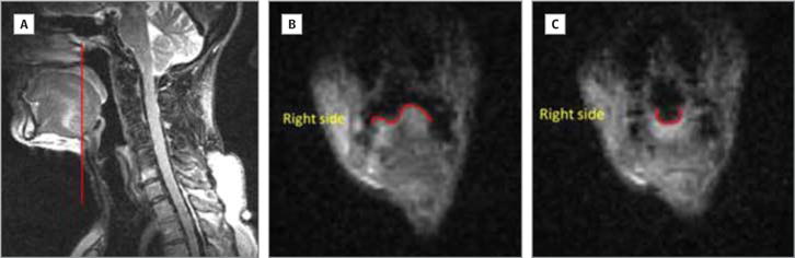
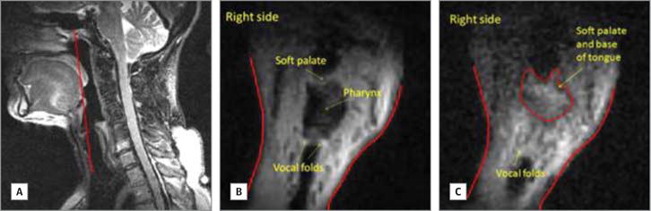
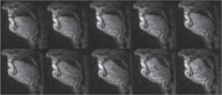
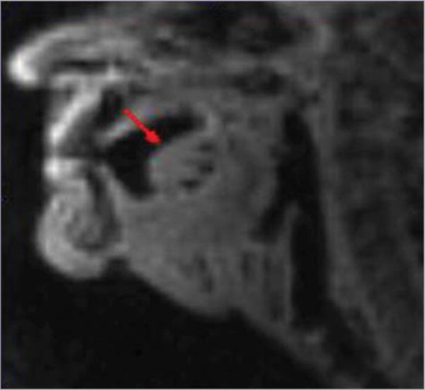
Interventions: Real-time MRI of the midsagittal plane was collected during swallowing. Coronal planes between the oral tongue and base of tongue and through the middle of the larynx were collected from 1 of the patients.
Main outcomes and measures: Oral transit time, pharyngeal transit time, submental muscle length change, and the distance change between the hyoid bone and anterior boundary of the thyroid cartilage were measured frame by frame during swallowing.
Results: All the measurable oral transit and pharyngeal transit times of the patients with cancer were significantly longer than the ones of the healthy participants. The changes in submental muscle length and the distance between the hyoid bone and thyroid cartilage happened in concert for all 60 normal swallows; however, the pattern differed for each patient with cancer. To our knowledge, the coronal view of the tongue and larynx revealed information that has not been previously reported.
Conclusions and relevance: This study has demonstrated the potential of real-time MRI to reveal critical information beyond the capacity of traditional videofluoroscopy. Further investigation is needed to fully consider the technique, procedure, and standard scope of applying MRI to evaluate swallow function of patients with cancer in research and clinic practice.
Conflict of interest statement
Disclosures: None reported.
Figures


References
-
- Warnakulasuriya S. Global epidemiology of oral and oropharyngeal cancer. Oral Oncol. 2009;45(4–5):309–316. - PubMed
-
- List MA, D’Antonio LL, Cella DF, et al. The performance status scale for head and neck cancer patients and the functional assessment of cancer therapy-head and neck scale. a study of utility and validity. Cancer. 1996;77(11):2294–2301. - PubMed
-
- Clark JR, Frei E., III Chemotherapy for head and neck cancer: progress and controversy in the management of patients with M0 disease. Semin Oncol. 1989;16(4 suppl 6):44–57. - PubMed
-
- Stoeckli SJ, Huisman TA, Seifert B, Martin-Harris BJ. Interrater reliability of videofluoroscopic swallow evaluation. Dysphagia. 2003;18(1):53–57. - PubMed
-
- Narayanan S, Nayak K, Lee S, Sethy A, Byrd D. An approach to real-time magnetic resonance imaging for speech production. J Acoust Soc Am. 2004;115(4):1771–1776. - PubMed
Publication types
MeSH terms
Grants and funding
LinkOut - more resources
Full Text Sources
Other Literature Sources
Medical
