

Neural substrates of inhibitory control deficits in 22q11.2 deletion syndrome
- PMID: 24177988
- PMCID: PMC4366617
- DOI: 10.1093/cercor/bht304
Neural substrates of inhibitory control deficits in 22q11.2 deletion syndrome
Abstract
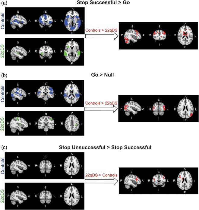
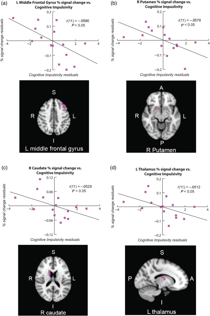
22q11.2 deletion syndrome (22q11DS) is associated with elevated levels of impulsivity, inattention, and distractibility, which may be related to underlying neurobiological dysfunction due to haploinsufficiency for genes involved in dopaminergic neurotransmission (i.e. catechol-O-methyltransferase). The Stop-signal task has been employed to probe the neural circuitry involved in response inhibition (RI); findings in healthy individuals indicate that a fronto-basal ganglia network underlies successful inhibition of a prepotent motor response. However, little is known about the neurobiological substrates of RI difficulties in 22q11DS. Here, we investigated this using functional magnetic resonance imaging while 45 adult participants (15 22q11DS patients, 30 matched controls) performed the Stop-signal task. Healthy controls showed significantly greater activation than 22q11DS patients within frontal cortical and basal ganglia regions during successful RI, whereas 22q11DS patients did not show increased neural activity relative to controls in any regions. Using the Barratt Impulsivity Scale, we also investigated whether neural dysfunction during RI was associated with cognitive impulsivity in 22q11DS patients. RI-related activity within left middle frontal gyrus and basal ganglia was associated with severity of self-reported cognitive impulsivity. These results suggest reduced engagement of RI-related brain regions in 22q11DS patients, which may be relevant to characteristic behavioral manifestations of the disorder.
Keywords: fronto-basal ganglia; impulsivity; response inhibition; velocardiofacial syndrome.
© The Author 2013. Published by Oxford University Press. All rights reserved. For Permissions, please e-mail: journals.permissions@oup.com.
Figures
References
-
- Alexander GE, Crutcher MD. Functional architecture of basal ganglia circuits: neural substrates of parallel processing. Trends Neurosci. 1990;13(7):266–271. - PubMed
-
- Antshel KM, Fremont W, Kates WR. The neurocognitive phenotype in velo-cardio-facial syndrome: a developmental perspective. Dev Disabil Res Rev. 2008;14:43–51. - PubMed
-
- Antshel KM, Stephen VF, Fremont W, Monuteaux MC, Kates WR, Doyle A, Mick E, Biederman J. Comparing ADHD in velocardiofacial syndrome to idiopathic ADHD: a preliminary study. J Atten Disord. 2007;11(1):64–73. - PubMed
-
- Aron AR, Poldrack RA. The cognitive neuroscience of response inhibition: relevance for genetic research in attention-deficit/hyperactivity disorder. Biol Psychiatry. 2005;57(11):1285–1292. - PubMed
Publication types
MeSH terms
Grants and funding
LinkOut - more resources
Full Text Sources
Other Literature Sources
