. 2015 Apr;25(4):1004-19.
doi: 10.1093/cercor/bht291. Epub 2013 Oct 31.

Ventral fronto-temporal pathway supporting cognitive control of episodic memory retrieval

Affiliations

Ventral fronto-temporal pathway supporting cognitive control of episodic memory retrieval

Jennifer Barredo et al. Cereb Cortex. 2015 Apr.

Abstract

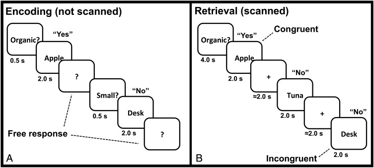
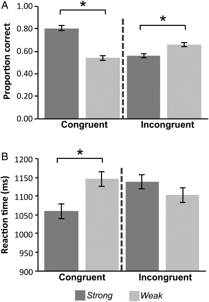
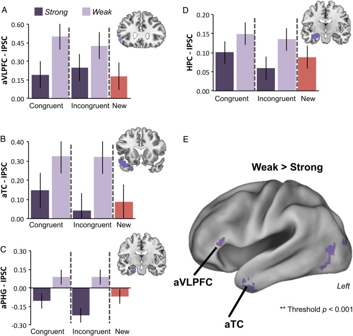
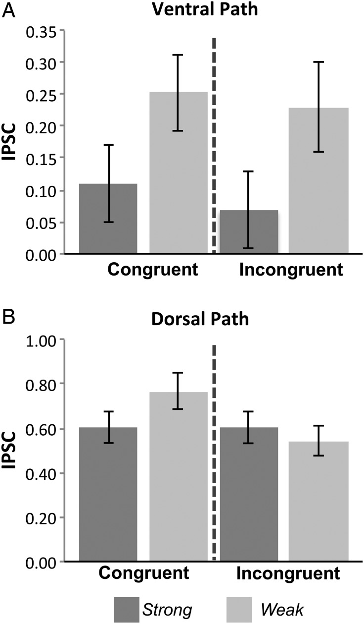
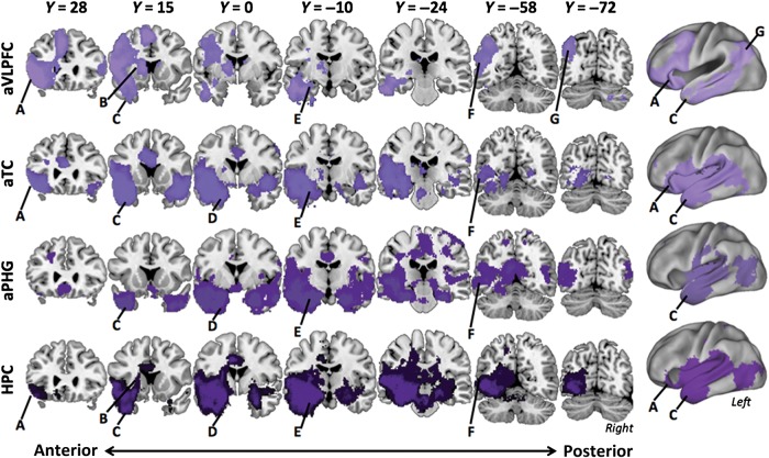
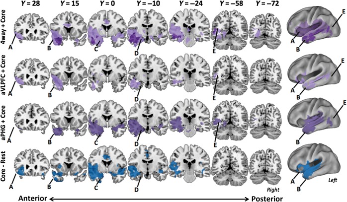
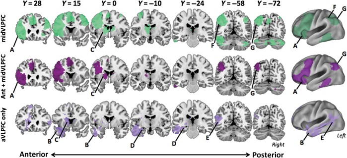
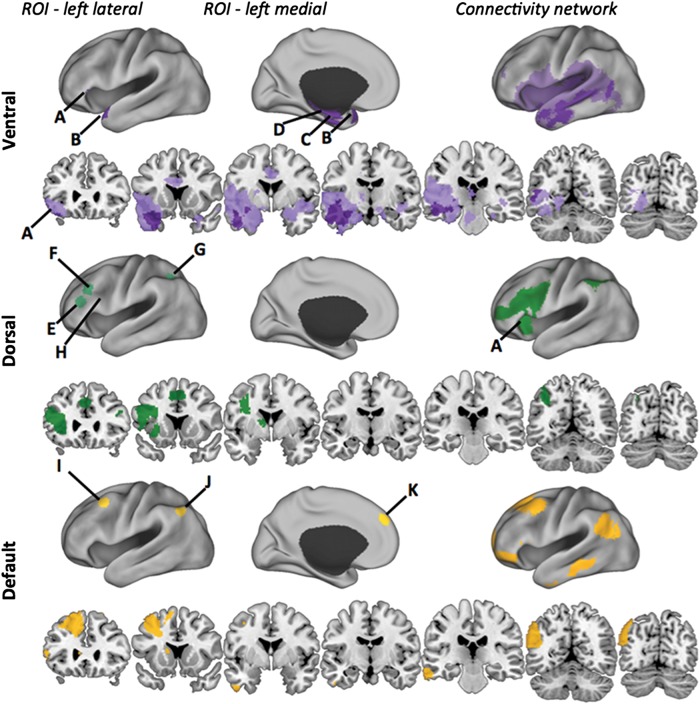
Achieving our goals often requires guiding access to relevant information from memory. Such goal-directed retrieval requires interactions between systems supporting cognitive control, including ventrolateral prefrontal cortex (VLPFC), and those supporting declarative memory, such as the medial temporal lobes (MTL). However, the pathways by which VLPFC interacts with MTL during retrieval are underspecified. Prior neuroanatomical evidence suggests that a polysynaptic ventral fronto-temporal pathway may support VLPFC-MTL interactions. To test this hypothesis, human participants were scanned using fMRI during performance of a source-monitoring task. The strength of source information was varied via repetition during encoding. Single encoding events should produce a weaker memory trace, thus recovering source information about these items should demand greater cognitive control. Results demonstrated that cortical targets along the ventral path--anterior VLPFC, temporal pole, anterior parahippocampus, and hippocampus--exhibited increases in univariate BOLD response correlated with increases in controlled retrieval demand, independent of factors related to response selection. Further, a functional connectivity analysis indicated that these regions functionally couple and are distinguishable from a dorsal pathway related to response selection demands. These data support a ventral retrieval pathway linking PFC and MTL.

Keywords: VLPFC; functional connectivity; retrieval.


Figures

Figure 1.
Figure 1.
Schematic of the trial events during the encoding and Source tasks. (A) During encoding, trials were initiated by pressing a keyboard space bar that prompted the display of a semantic decision cue (0.5 s). This was followed by the display of a word (2 s), followed by a prompt (?) that remained on the screen until a response was made. Participants had unlimited time to answer “Yes” or “No” for that item with respect to the orienting question and the next trial cycle did not begin until it was initiated by the participant. (B) During source retrieval blocks, participants reported whether presented words were encountered at encoding with the orienting question displayed at block start. Target source task prompts (either “Organic?” or “Small?”) were displayed for 4 s at the start of each block, and were followed by a 12 s baseline and then word targets were presented for 2 s each. Participants responded “Yes” or “No” during the 2 s target presentation. A jittered ITI separated retrieval trials within blocks.
Figure 2.
Figure 2.
Behavioral results (Accuracy and RT) for Congruency and Strength conditions of the Source task. Congruency and Strength produced a crossover interaction in Accuracy and RT, such that Weak Congruent trials were associated with lower accuracy and slower RT than Strong Congruent, but Strong Incongruent produced worse performance than Weak Incongruent. Error bars depict within-subject standard error (*P < 0.05).
Figure 3.
Figure 3.
Effects of Strength and Congruency along the ventral pathway. Bars plot IPSC from the crossing of Strength and Congruency in the ROI analyses of the a priori ventral path regions: (A) aVLPFC, (B) aTC, (C) aPHG, and (D) HPC. Response to New items is also plotted for reference. In general, IPSC tracks Strength independently of Congruency in these regions consistent with a controlled retrieval process. Error bars depict within-subject standard error. (E) Surface rendering on the left hemisphere of the whole-brain voxelwise contrast of Weak > Strong (P < 0.001, uncorrected). This contrast shows that the effects of Strength are primarily in aVLPFC and temporal/ventral occipital cortex. The VLPFC activation cluster straddles the horizontal ramus of the lateral fissure, and so includes dorsal portions of BA47 and the rostral portion of BA45. Error bars depict within-subject standard error.
Figure 4.
Figure 4.
The ventral and dorsal pathways are involved in separable control processes. Activation in regions defined a priori along the ventral path (top) track the modulation of Strength rather than Congruency or the interaction of both factors. Importantly, significantly greater activation is observed for Weak than Strong within the Incongruent condition (P < 0.05), a pattern inconsistent with a response selection effect. In contrast, activation in regions defined a priori along the fronto-parietal “dorsal path” (bottom) reliably track the interaction of Strength and Congruency, implicating this pathway in postretrieval selection rather than memory access. Error bars depict within-subject standard error.
Figure 5.
Figure 5.
Functional connectivity along the ventral path during Source retrieval. Voxelwise connectivity of each of the 4 ventral path seeds (labels at left of each row) is plotted. Functional networks across Weak and Strong conditions were similar within each of these seeds, thus only the Weak Source condition is pictured. Major structures of interest are labeled: (A) aVLPFC, (B) striatum, (C) aTC, (D) aPHG, (E) HPC, (F) cMTG, (G) angular gyrus/IPS.
Figure 6.
Figure 6.
Conjunction analyses of connectivity along the ventral pathway. Voxelwise results from 4 connectivity analyses (labels at left of each row) are plotted on coronal slices. All contrasts are valid “AND” conjunctions from FDR-corrected seed maps thresholded at P < 0.05. Major structures of interest are labeled: (A) aVLPFC, (B) aTC, (C) aPHG, (D) HPC, and (E) cMTG. The top row plots conjunction analyses showing the most (light plot) and least (dark plot) inclusive definitions of the retrieval network. The aTC–HPC connectivity conjunction network is in light purple, and the more restricted the 4-way conjunction of the connectivity maps from the 4 a priori seeds is plotted in dark purple. The second and third rows depict the aVLPFC-conjunction in lilac (labeled “aVLPFC Conj.”) and aPHG-conjunction in medium purple (labeled “aPHG Conj.”), respectively. The bottom row (labeled “Core–rest”) plots results of the aTC–HPC conjunction at rest in blue.
Figure 7.
Figure 7.
Analyses of specificity of the ventral path network. (A) Voxelwise results from the functional connectivity analysis of the mid-VLPFC seed are plotted in green. Mid-VLPFC includes mostly dorsal frontal-parietal sites with aVLPFC being the only ventral path region in its network (<10 voxels, (A). AVLPFC and mid-VLPFC also share voxels in mid-caudal MTG, but shared voxels are sparse and are not easily visualized. (B) The conjunction (P < 0.05 FDR) of the mid-VLPFC and aVLPFC connectivity maps reveals overlap among the dorsolateral PFC and parietal components of the aVLPFC network (maroon). (C) The subtraction of the connectivity map of mid-VLPFC from aVLPFC reveals regions of the ventral retrieval network plotted in lilac.
Figure 8.
Figure 8.
Comparison of default and fronto-parietal control network seeds to the retrieval network. The ventral path connectivity network is plotted in purple (top), with the lighter shade denoting the Core connectivity network and the darker the 4-way conjunction. ROIs are defining ventral path network are marked and include (A) aVLPFC, (B) aTC, (C) aPHG, and (D) HPC. The dorsal network is plotted in green (middle). The fronto-parietal ROIs are marked and include (E) PFCl, (F) PFClp, (G) IPS, and (H) mid-VLPFC. Note that mid-VLPFC lies within the sulcus and cannot be seen in lateral view; approximate location only is marked. The conjunction of the networks generated from 3 a priori default network seeds, (I) PFCdp, (J) PGpd, and (K) PFCdm, are plotted in yellow (bottom). Comparison across networks illustrates that the overlap between fronto-parietal control network and retrieval network is primarily limited to aVLPFC. The retrieval network overlaps with the ventral portion of the default network.

References

    1. Badre D, Poldrack R, Paré-Blagoev EJ, Insler RZ, Wagner AD. Dissociable controlled retrieval and generalized selection mechanisms in ventrolateral prefrontal cortex. Neuron. 2005;47(6):907–918. - PubMed
    1. Badre D, Wagner AD. Computational and neurobiological mechanisms underlying cognitive flexibility. Proc Natl Acad Sci U S A. 2006;103(18):7186–7191. - PMC - PubMed
    1. Badre D, Wagner AD. Left ventrolateral prefrontal cortex and the cognitive control of memory. Neuropsychologia. 2007;45(13):2883–2901. - PubMed
    1. Benjamin AS. Memory is more than just remembering: strategic control of encoding, accessing memory, and making decisions. In: Benjamin AS, Ross BH, editors. The psychology of learning and motivation: skill and strategy in memory use. Vol. 48. London: Academic Press; 2007. pp. 1–71.
    1. Blaizot X, Mansilla F, Insausti AM, Constans JM, Salinas-Alamán A, Pró-Sistiaga P, Mohedano-Moriano A, Insausti R. The human parahippocampal region: I. Temporal pole cytoarchitectonic and MRI correlation. Cereb Cortex. 2010;20(9):2198–2212. - PMC - PubMed

Publication types