. 2014 Feb;42(3):1553-62.
doi: 10.1093/nar/gkt968. Epub 2013 Oct 30.

A pathogenic non-coding RNA induces changes in dynamic DNA methylation of ribosomal RNA genes in host plants

Affiliations

A pathogenic non-coding RNA induces changes in dynamic DNA methylation of ribosomal RNA genes in host plants

German Martinez et al. Nucleic Acids Res. 2014 Feb.

Abstract

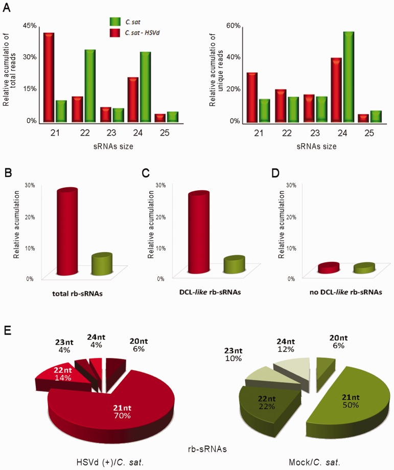
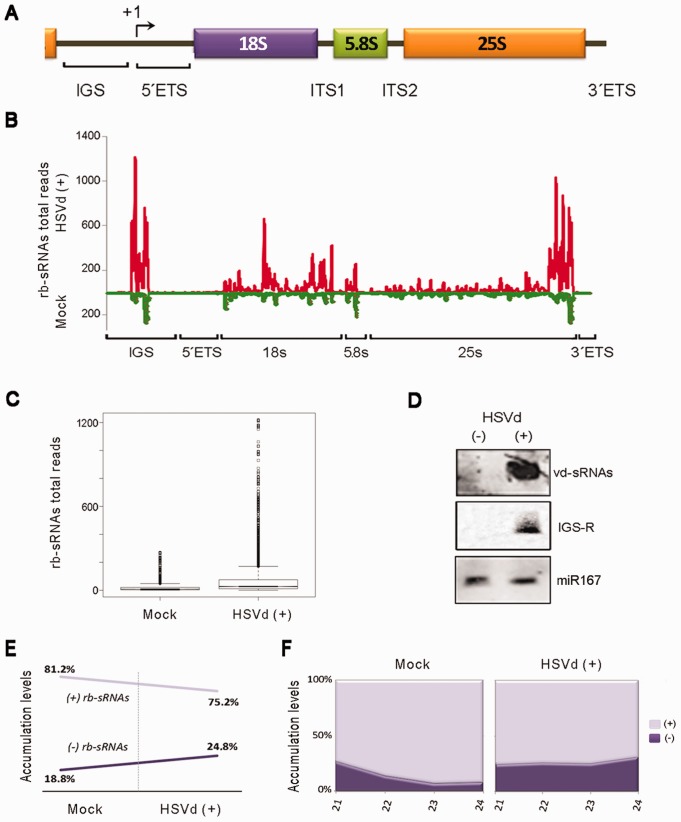
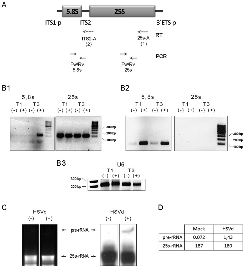
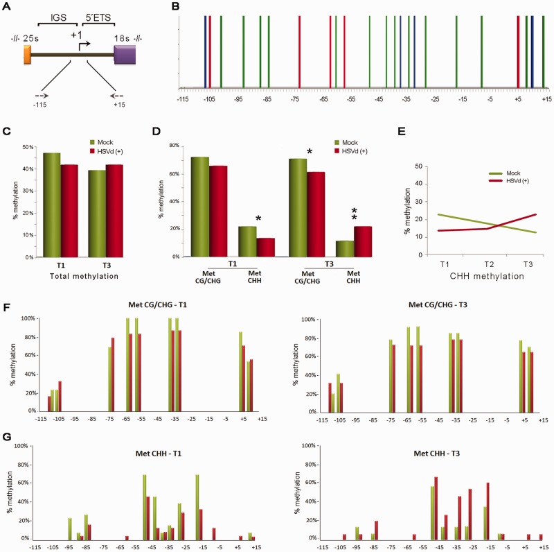
Viroids are plant-pathogenic non-coding RNAs able to interfere with as yet poorly known host-regulatory pathways and to cause alterations recognized as diseases. The way in which these RNAs coerce the host to express symptoms remains to be totally deciphered. In recent years, diverse studies have proposed a close interplay between viroid-induced pathogenesis and RNA silencing, supporting the belief that viroid-derived small RNAs mediate the post-transcriptional cleavage of endogenous mRNAs by acting as elicitors of symptoms expression. Although the evidence supporting the role of viroid-derived small RNAs in pathogenesis is robust, the possibility that this phenomenon can be a more complex process, also involving viroid-induced alterations in plant gene expression at transcriptional levels, has been considered. Here we show that plants infected with the 'Hop stunt viroid' accumulate high levels of sRNAs derived from ribosomal transcripts. This effect was correlated with an increase in the transcription of ribosomal RNA (rRNA) precursors during infection. We observed that the transcriptional reactivation of rRNA genes correlates with a modification of DNA methylation in their promoter region and revealed that some rRNA genes are demethylated and transcriptionally reactivated during infection. This study reports a previously unknown mechanism associated with viroid (or any other pathogenic RNA) infection in plants providing new insights into aspects of host alterations induced by the viroid infectious cycle.


Figures

Figure 1.
Figure 1.
Characterization of the sRNAs recovered from cucumber leaves by deep sequencing. (A) Graphic representation of the differential accumulation and distribution of the total (left) and unique (right) reads of endogenous cucumber sRNAs ranging between 21 and 25 nt recovered from both the control and HSVd-infected samples analyzed at 30 days post-inoculation. (B) Differential recovering of rb-sRNAs from infected and healthy tissues. The accumulation of rb-sRNAs is expressed as the percentage of total rb-sRNAs from the overall sRNAs in the library. (C) and (D) show the relative accumulation of recovered rb-sRNAs with canonical (21–24 nt) and non-canonical (<21 and >24 nt) predicted sizes, respectively. (E) Graphic representation of the distribution of the total reads of rb-sRNAs (ranging between 21 and 25 nt) recovered from both the HSVd-infected (left) and control (right) analyzed samples.
Figure 2.
Figure 2.
Analysis of ribosomal-derived sRNAs differentially expressed in infected cucumber plants. (A) Diagram (no scale) of the rRNA gene unit. The rRNA gene repeats are arranged in long tandem arrays of 45S rRNA genes, each including the region for the 18S, 5.8S and 25S rRNAs, and separated from adjacent units by an IGS. The transcription start site is indicated by +1. The ETS and ITS are removed during rRNA processing. (B) The rb-sRNAs recovered from the infected (above the X-axis) or the non-inoculated plants (below the X-axis) were plotted according to the position of their 5′-end onto the cucumber rRNA sequence. The values on the Y-axis represent the number of total reads in each library. The nucleotide positions −115 to +15 of the 45S rRNA-analyzed region are represented on the X-axis. (C) Comparison of the accumulation of the rb-sRNAs that map along the rRNA transcriptional unit between both the infected and control plants as shown in the box-plot (boxes represent the medians, and the first and third quartiles of the dataset; circles refer to the data whose values are beyond the quartiles. Comparison of the means of the boxes showed significant differences between both samples using a parametric t-test: arithmetic means for mock and infected plants are 9.49 and 37.66, respectively; t = 27.11; P < 2.2 × 10⁻¹⁶). (D) The hyper-accumulation in the infected samples of the rb-sRNAs derived from an ∼300-nt region of the IGS (IGS-R) was validated by northern blot assays. The miR167 equally recovered from both analyzed sRNA datasets was used as a load control. Hybridization with a probe against the HSVd-derived sRNAs (vd-sRNAs) was used as a positive control. (E) The relative accumulation of total rb-sRNAs complementary to rRNA transcripts increased in the HSVd-infected plants (left). A similar result was obtained when individually analyzing the expected canonical size of the rb-sRNAs (right).
Figure 3.
Figure 3.
Precursor for rRNAs (pre-rRNAs) accumulates in infected plants. (A) Diagram (no scale) of the pre-27S rRNA (ITS1-p and 3′ETS-p refer to partially processed transcribed spacers). The dotted arrows below depict the oligos used for the RT reaction starting from the 25S 3′-end (25s-A/RT-1) and the ITS2 (ITS2-A/RT-2) regions of the pre-rRNA. Solid arrows indicate the oligos used in the PCR amplification. (B) The RT-PCR analyses of the pre-rRNA expression in the HSVd-infected (+) and mock-inoculated (−) plants at 10 (T1) and 30 (T3) days post-inoculation. The initial cDNAs were transcribed using the 25s-A (B1) and ITS2-A (B2) oligos, respectively. (B3) RT-PCR amplification of U6 Small nucleolar RNA (snoRNA) served as control for RNA load. (C) The accumulation of the pre-rRNA (marked with arrows) in the infected plants was validated by northern blot assays in the total RNAs extracted from plants at T3 using a probe complementary to the 3′-end of the 25S rRNA. (D) The band intensity was measured using the Image-J application http://www.imagej.en.softonic.com.
Figure 4.
Figure 4.
HSVd infection affects the methylation patterns in 45S rRNA genes. (A) Diagram of the rRNA gene intergenic region highlighting the promoter zone analyzed by bisulfite sequencing. The arrows represent the oligos used in the PCR assay and their relative position in the rRNA gene. (B) Graphic representation of the potential symmetric (CG–red bars and CHG–blue bars) and asymmetric (CHH–green bars) positions predicted to exist within the analyzed region. (C) Histogram documenting the relative (HSVd/mock) total DNA methylation levels at infection times T1 and T3. (D) Schematic representation of the differential analysis of both symmetric and asymmetric cytosine methylation at infection times T1 and T3 (paired t-test values T1: means symmetric methylation (mock) 0.73, (infected) 0.67, t = 1.604; means CHH methylation (mock) 0.22, (infected) 0.13, t = 2.180; T3: means symmetric methylation (mock) 0.72, (infected) 0.64, t = 2.481; means CHH methylation (mock) 0.11, (infected) 0.22, t = 3.047; *P < 0.05, **P < 0.01). (E) Evolution of the CHH methylation during HSVd infection in comparison with the level observed in the mock-inoculated plants. (F) Positions of methylcytosines in the analyzed regions displayed in the symmetric (CG and CHG) context. (G) Positions of methylcytosines in the analyzed regions displayed in the asymmetric context. The height of the bar represents the frequency at which cytosine was methylated at the analyzed infection times T1 (left) and T3 (right).

Similar articles

Cited by

References

    1. Ding B. The biology of viroid-host interactions. Annu. Rev. Phytopathol. 2009;47:105–131. - PubMed
    1. Ding B. Viroids: self-replicating, mobile, and fast-evolving noncoding regulatory RNAs. Wiley Interdiscip. Rev. RNA. 2010;1:362–375. - PubMed
    1. Navarro B, Gisel A, Rodio ME, Delgado S, Flores R, Di Serio F. Viroids: how to infect a host and cause disease without encoding proteins. Biochimie. 2012;94:1474–1480. - PubMed
    1. Zhao Y, Owens RA, Hammond RW. Use of a vector based on potato virus X in a whole plant assay to demonstrate nuclear targeting of potato spindle tuber viroid. J. Gen. Virol. 2001;82:1491–1497. - PubMed
    1. Gomez G, Pallas V. Studies on subcellular compartmentalization of plant pathogenic noncoding RNAs give new insights into the intracellular RNA-traffic mechanisms. Plant Physiol. 2012;159:558–564. - PMC - PubMed

Publication types

Associated data