. 2013 Dec 20;288(51):36610-23.
doi: 10.1074/jbc.M113.453183. Epub 2013 Oct 31.

Proprotein convertase subtilisin/kexin type 7 (PCSK7) is essential for the zebrafish development and bioavailability of transforming growth factor β1a (TGFβ1a)

Affiliations

Proprotein convertase subtilisin/kexin type 7 (PCSK7) is essential for the zebrafish development and bioavailability of transforming growth factor β1a (TGFβ1a)

Hannu Turpeinen et al. J Biol Chem. .

Abstract

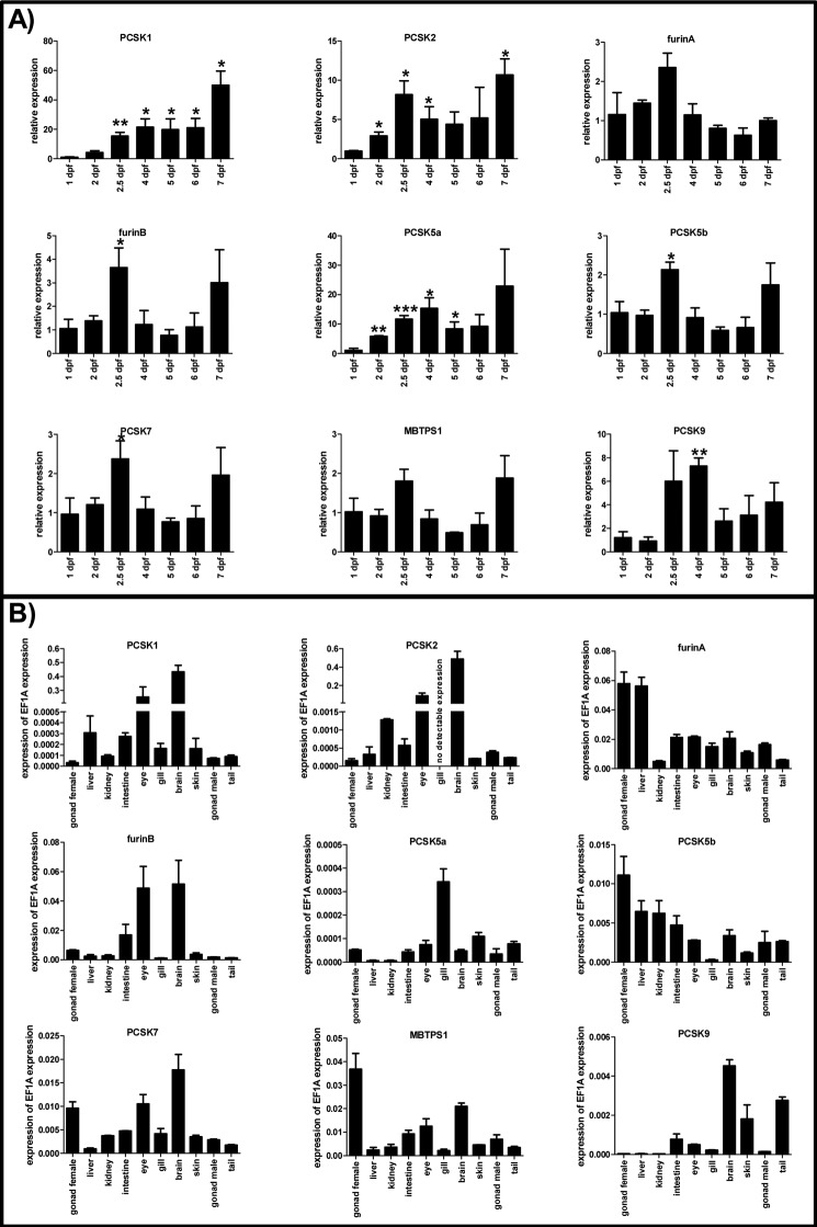
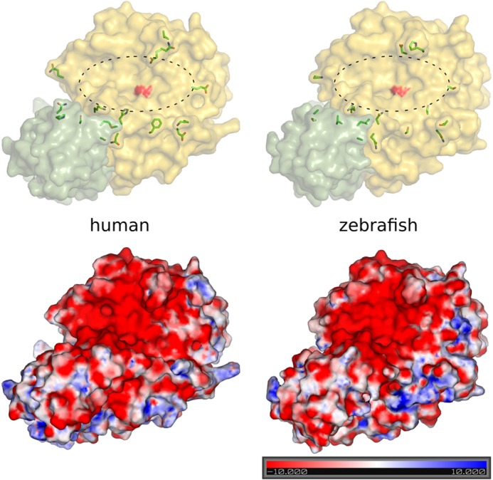
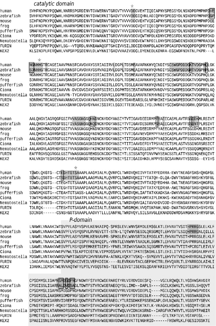
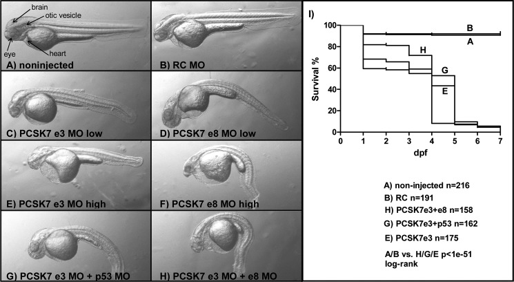
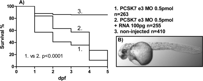
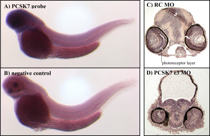
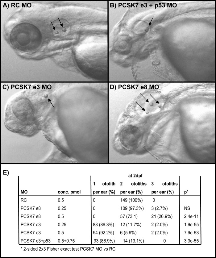
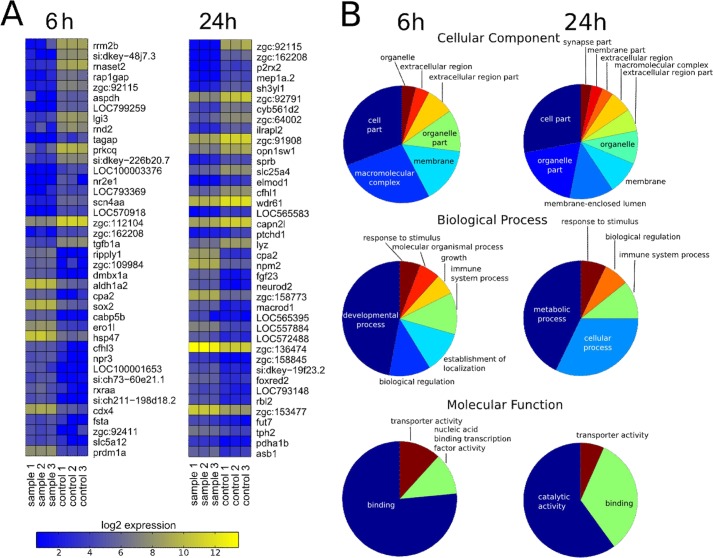
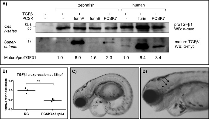
Proprotein convertase subtilisin/kexin (PCSK) enzymes convert proproteins into bioactive end products. Although other PCSK enzymes are known to be essential for biological processes ranging from cholesterol metabolism to host defense, the in vivo importance of the evolutionarily ancient PCSK7 has remained enigmatic. Here, we quantified the expressions of all pcsk genes during the 1st week of fish development and in several tissues. pcsk7 expression was ubiquitous and evident already during the early development. To compare mammalian and zebrafish PCSK7, we prepared homology models, which demonstrated remarkable structural conservation. When the PCSK7 function in developing larvae was inhibited, we found that PCSK7-deficient fish have defects in various organs, including the brain, eye, and otic vesicle, and these result in mortality within 7 days postfertilization. A genome-wide analysis of PCSK7-dependent gene expression showed that, in addition to developmental processes, several immune system-related pathways are also regulated by PCSK7. Specifically, the PCSK7 contributed to the mRNA expression and proteolytic cleavage of the cytokine TGFβ1a. Consequently, tgfβ1a morphant fish displayed phenotypical similarities with pcsk7 morphants, underscoring the importance of this cytokine in the zebrafish development. Targeting PCSK activity has emerged as a strategy for treating human diseases. Our results suggest that inhibiting PCSK7 might interfere with normal vertebrate development.

Keywords: Development; Gene Expression; Homology Modeling; Otolith; PC7; PCSK7; Proprotein Convertase; Protease; Transforming Growth Factor β (TGFβ); Zebrafish.


Figures

FIGURE 1.
FIGURE 1.
Expression of pcsk genes in zebrafish. QRT-PCR analysis was used to analyze the relative expression of the pcsk genes in developing zebrafish larvae (1–7 dpf) (A) and various adult zebrafish tissues (B). A, pcsk gene expression levels were normalized to the housekeeping gene ef1a, and the normalized gene expression on 1 dpf was given a value of 1. Other time points are shown as relative to this. Asterisks denote statistical significance for differences in comparisons between 1 dpf and other time points: *, p < 0.05; **, p < 0.01; ***, p < 0.001 in Welch-corrected two-tailed Student's t tests. Experiments in A were performed with three biological replicates (each sample consisted of 15–30 individual larvae depending on the age of larvae). B, in adult tissue analyses, gene expression levels related to that of ef1a are shown. Experiments in B were performed twice in technical replicates with essentially similar results. Error bars represent S.D. Note the diverse scales on the y axis.
FIGURE 2.
FIGURE 2.
Homology models of human and zebrafish PCSK7. Upper part, yellow, catalytic domain; green, P domain. The catalytic site is marked with an oval, and the surface of the catalytic serine is highlighted in red. Amino acid differences close to the conserved substrate binding site are shown in stick presentation. Lower part, electrostatic potentials (kT/e) for the models were calculated using APBS 1.3 (57) and visualized in PyMOL 2.7. Red-white-blue color indicates the ±10 kT/e electrostatic potential plotted on the protein surface.
FIGURE 3.
FIGURE 3.
ClustalW alignment of PCSK7 homologs. Human (Homo sapiens, UniProt Q16549), zebrafish (Danio rerio, RefSeq NP_001076494.1), mouse (Mus musculus, UniProt Q61139), frog (Xenopus laevis, RefSeq NP_001090019.1), pufferfish (Tetraodon nigroviridis, Ensembl ENSTNIP00000017367), vase tunicate (Ciona intestinalis, RefSeq XP_002125956.1), and starlet sea anemone (Nematostella vectensis, RefSeq XP_001638665.1) PCSK7 catalytic and P domains were aligned with those of mouse FURIN (Protein Data Bank code 1P8J (58)) and yeast Kexin (Saccharomyces cerevisiae, Protein Data Bank code 2ID4 (59)). The catalytic triad is marked with stars, and potential substrate-binding residues are in dark gray. Residues showing differences in Fig. 2 are highlighted with rectangles.
FIGURE 4.
FIGURE 4.
Phenotypes and survival of pcsk7 morphant fish. Zebrafish larvae (2 dpf) were non-injected (A) or injected with RC MO (0.5 pmol) (B), pcsk7 e3 MO (0.25 pmol) (C), pcsk7 e8 MO (0.25 pmol) (D), pcsk7 e3 MO (0.5 pmol) (E), pcsk7 e8 MO (0.5 pmol) (F), pcsk7 e3 + p53 MO (0.5 + 0.75 pmol) (G), or pcsk7 e3 + e8 MO (0.25 + 0.25 pmol) (H). A–H, 35× magnification. I, survival of fish injected with pcsk7 e3 MO (0.5 pmol), pcsk7 e3 + e8 MOs (0.25 + 0.25 pmol), and pcsk7 e3 + p53 MOs (0.5 + 0.75 pmol) was significantly lower than that of RC morphant (0.5 pmol) or uninjected fish (p < 1e−51 for all comparisons by log rank test). pcsk7 e8 MO reduced the survival of larvae similarly to e3 MO: all larvae died by 6 dpf (n = 98 larvae; e8 MO, 0.5 pmol; data not shown).
FIGURE 5.
FIGURE 5.
pcsk7 mRNA improves the survival and reduces the severity of the pcsk7 morphant phenotype. A, survival of zebrafish uninjected or injected with pcsk7 e3 MO alone or together with in vitro transcribed pcsk7 mRNA was monitored daily. Data are pooled from two independent experiments. B, 2-dpf zebrafish larvae co-injected with pcsk7 e3 MO (0.5 pmol) and pcsk7 mRNA (100 pg).
FIGURE 6.
FIGURE 6.
pcsk7 MO injections result in erratic splicing of the pre-mRNA. A, total RNA was isolated from the control (RC MO) and different pcsk7 morphant fish and reverse transcribed into cDNA, which was amplified by PCR and run on an agarose gel. From the left, lane 1, 100-bp molecular weight marker; lane 2, pcsk7 e3 MO; lane 3, control (=RC MO with pcsk7 e3 primers); lane 4, pcsk7 e8 MO; lane 5, control (=RC MO with pcsk7 e8 primers); lane 6, 100-bp molecular weight marker. Sequencing showed that 345- and 288-bp DNA fragments represent intact, wild-type pcsk7 mRNA that can be detected in RC MO samples with the e3 (lane 3) and e8 primers (lane 5), respectively. The pcsk7 e3 MO injection resulted in three differentially sized fragments: (i) a fragment corresponding to the completely deleted exon 3 (the shortest band in lane 2), (ii) a fragment with a 58-bp deletion from the start of exon 3 followed by a 49-bp polymorphic region either from exon 3 or from exon 4 and further supplemented with the untouched end of exon 3 (the middle band in lane 2), and (iii) a faint band of wild-type exon 3 (the longest band in lane 2). The pcsk7 e8 MO injection deleted a 58-bp fragment from the end of exon 8 (lane 4) resulting in a truncated pcsk7 mRNA molecule. B, sequences for the primers used for the sequencing described in A. F, forward; R, reverse.
FIGURE 7.
FIGURE 7.
pcsk7 is expressed in the head region, and pcsk7 morphant fish have cranial developmental defects. pcsk7 expression (dark blue) was analyzed with RNA in situ hybridization in wild-type zebrafish larvae at 2 dpf. A, pcsk7 antisense probe. B, negative control (pcsk7 sense probe). Right-hand panels show hematoxylin-eosin-stained transverse sections of zebrafish cranial region of RC (C) and pcsk7 e3 morphant (D) (3 dpf).
FIGURE 8.
FIGURE 8.
pcsk7 morphant fish have an abnormal number of otoliths. Zebrafish were injected with RC MO (0.5 pmol) (A), pcsk7 e3 + p53 MO (0.5 + 0.75 pmol) (B), pcsk7 e3 MO (0.5 pmol) (C), or pcsk7 e8 MO (0.5 pmol) (D), and otoliths (arrows) were visualized on 3 dpf. Quantification of otoliths is presented in E. NS, not significant.
FIGURE 9.
FIGURE 9.
Heat map of the most differentially expressed genes and high level summary of gene ontology enrichments between control and pcsk7 morphant fish. Samples were prepared, and data were analyzed as described under “Experimental Procedures.” A, zebrafish genes that have an identifiable human homolog are shown in the heat maps. B, summaries from the enriched gene ontology terms. Pie charts show enriched high level categories for cellular component, biological process, and molecular function at 6 and 24 hpf. In each chart, the size of the wedge corresponds to the number of terms enriched under the given high level category.
FIGURE 10.
FIGURE 10.
PCSK7 regulates TGFβ1a, which affects the zebrafish larva development and otolith formation. A, FURIN-deficient RPE.40 cells were transiently transfected with zebrafish tgfβ1a-myc or human TGFβ1-myc together with zffurinA, zffurinB, zfpcsk7, huFURIN, or huPCSK7. Pro-TGFβ1 (45 kDa) and mature TGFβ1 (16 kDa in zebrafish and 14 kDa in human) expressions were detected with Western blotting (WB). Mature/pro-TGFβ1 ratios were quantified using NIH ImageJ software, and ratios in cells transfected only with tgfβ1 cDNAs were given an arbitrary value of 1. Equal loading of cell lysates and supernatants was verified by Ponceau S staining (data not shown). The experiment was repeated twice with similar results. B, tgfβ1a mRNA expression was measured by QRT-PCR from pcsk7 e3 + p53 and RC morphant embryos at 48 hpf (**, p = 0.0052, two-tailed Student's t test). C and D depict the phenotypes (4 dpf) of zebrafish injected with tgfβ1a + p53 MOs (tgfβ1a MO, 1.0 pmol; p53 MO, 1.5 pmol). tgfβ1a morphants had incorrect otolith numbers: tgfβ1a MO, 65 fish with two otoliths per ear, 21 fish with three otoliths per ear; RC MO, 120 fish with two otoliths per ear, 0 fish with three otoliths per ear (p = 2.16e−9 in Fisher 2 × 2 test).

Similar articles

Cited by

References

    1. Seidah N. G., Prat A. (2012) The biology and therapeutic targeting of the proprotein convertases. Nat. Rev. Drug Discov. 11, 367–383 - PubMed
    1. Roebroek A. J., Umans L., Pauli I. G., Robertson E. J., van Leuven F., Van de Ven W. J., Constam D. B. (1998) Failure of ventral closure and axial rotation in embryos lacking the proprotein convertase Furin. Development 125, 4863–4876 - PubMed
    1. Essalmani R., Hamelin J., Marcinkiewicz J., Chamberland A., Mbikay M., Chrétien M., Seidah N. G., Prat A. (2006) Deletion of the gene encoding proprotein convertase 5/6 causes early embryonic lethality in the mouse. Mol. Cell. Biol. 26, 354–361 - PMC - PubMed
    1. Constam D. B., Robertson E. J. (2000) SPC4/PACE4 regulates a TGFβ signaling network during axis formation. Genes Dev. 14, 1146–1155 - PMC - PubMed
    1. Zhu X., Zhou A., Dey A., Norrbom C., Carroll R., Zhang C., Laurent V., Lindberg I., Ugleholdt R., Holst J. J., Steiner D. F. (2002) Disruption of PC1/3 expression in mice causes dwarfism and multiple neuroendocrine peptide processing defects. Proc. Natl. Acad. Sci. U.S.A. 99, 10293–10298 - PMC - PubMed

Publication types

MeSH terms