. 2014 Jan;24(1):142-53.
doi: 10.1101/gr.161638.113. Epub 2013 Oct 31.

Highly efficient CRISPR/Cas9-mediated knock-in in zebrafish by homology-independent DNA repair

Affiliations

Highly efficient CRISPR/Cas9-mediated knock-in in zebrafish by homology-independent DNA repair

Thomas O Auer et al. Genome Res. 2014 Jan.

Abstract

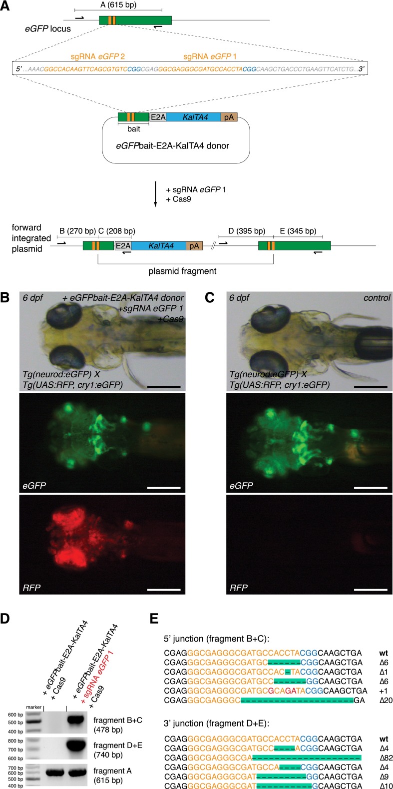
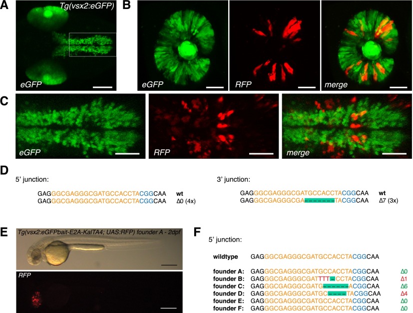
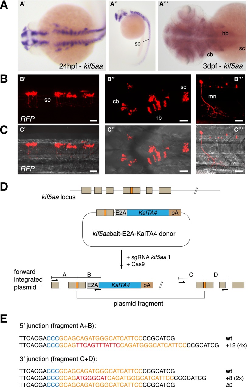
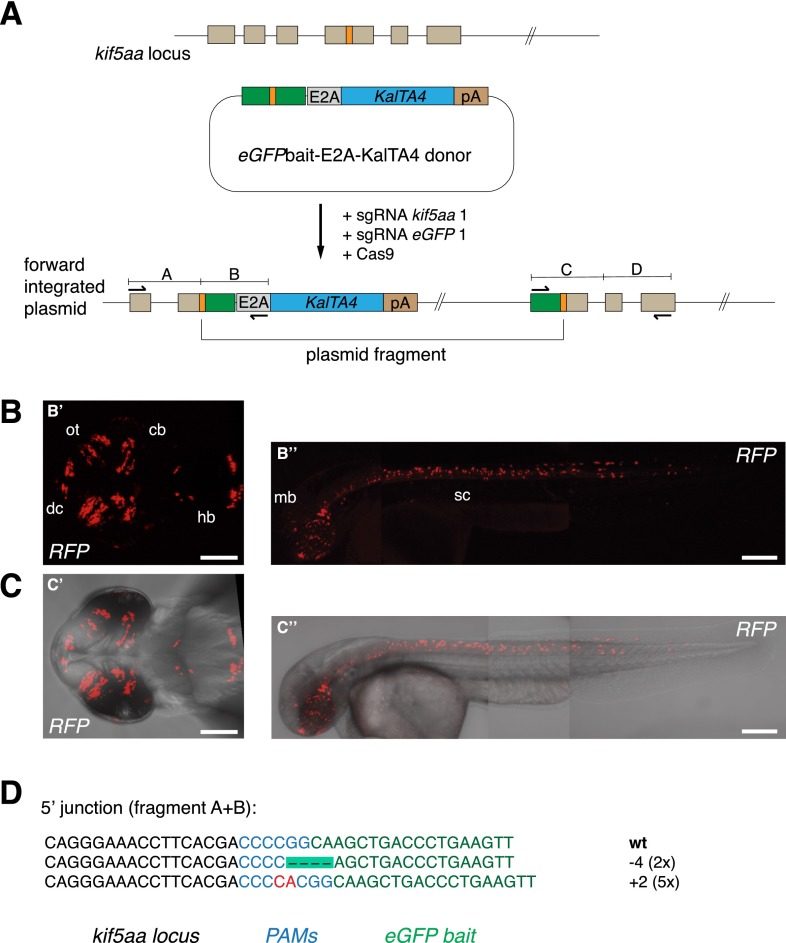
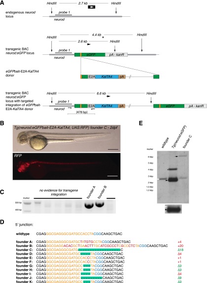
Sequence-specific nucleases like TALENs and the CRISPR/Cas9 system have greatly expanded the genome editing possibilities in model organisms such as zebrafish. Both systems have recently been used to create knock-out alleles with great efficiency, and TALENs have also been successfully employed in knock-in of DNA cassettes at defined loci via homologous recombination (HR). Here we report CRISPR/Cas9-mediated knock-in of DNA cassettes into the zebrafish genome at a very high rate by homology-independent double-strand break (DSB) repair pathways. After co-injection of a donor plasmid with a short guide RNA (sgRNA) and Cas9 nuclease mRNA, concurrent cleavage of donor plasmid DNA and the selected chromosomal integration site resulted in efficient targeted integration of donor DNA. We successfully employed this approach to convert eGFP into Gal4 transgenic lines, and the same plasmids and sgRNAs can be applied in any species where eGFP lines were generated as part of enhancer and gene trap screens. In addition, we show the possibility of easily targeting DNA integration at endogenous loci, thus greatly facilitating the creation of reporter and loss-of-function alleles. Due to its simplicity, flexibility, and very high efficiency, our method greatly expands the repertoire for genome editing in zebrafish and can be readily adapted to many other organisms.


Figures

Figure 1.
Figure 1.
CRISPR/Cas9-mediated knock-in of KalTA4 into the Tg(neurod:eGFP) transgenic line. (A) A schematic of the donor plasmid consisting of an N-terminal eGFPbait with two sgRNA target sites (in orange, PAM sequence in blue). After co-injection of the donor with Cas9 mRNA and one eGFP sgRNA, insertion at the eGFP locus occurs. In-frame fusion of the E2A-KalTA4-pA cassette results in a multicistronic mRNA after successful integration at the eGFP locus. Due to the E2A sequence, the N-terminal eGFP peptide is cleaved from the KalTA4 protein by cotranslational ribosomal skipping. (B) A 6-dpf Tg(neurod:eGFP) × Tg(UAS:RFP, cry1:eGFP) embryo showing a switch from eGFP- to RFP-expressing cells upon injection of the donor plasmid together with sgRNA eGFP 1 and Cas9 mRNA. Successful in-frame knock-in of the donor plasmid into the eGFP open reading frame results in KalTA4 expression. Consecutively, KalTA4 binds to UAS:RFP and triggers RFP expression, leading to the eGFP to RFP switch. Scale bar, 300 μm. Tg(UAS:RFP, cry1:eGFP) transgenic fish express eGFP in the lens (driven by the crystalline promoter cry1:eGFP), thus allowing UAS:RFP transgenic fish to be identified by expression of eGFP in their lens (since without transactivation by KalTA4, no RFP is expressed from this transgene). (C) No RFP-expressing cells could be observed in Tg(neurod:eGFP) × Tg(UAS:RFP, cry1:eGFP) embryos injected with the donor plasmid and Cas9 mRNA but without sgRNA eGFP 1. Scale bar, 300 μm. (D) A representative gel of PCR products obtained from the founder fish shown in B, demonstrating targeted knock-in of the donor plasmid at the eGFP locus. PCR primers were placed flanking the neurod:eGFP locus and outward directed in the donor plasmid. Positions of PCR primers and the resulting fragment nomenclature are shown in A. (E) Sequence analysis at the 5′ and 3′ junctions of five representative targeted integration events. (Orange) sgRNA binding site, (red) base pair changes or insertions. The PAM sequence NGG required for cleavage by Cas9 (Jinek et al. 2012) is shown in blue. Note that only the Δ6 integration events correspond to in-frame insertions of the E2A-KalTA4 sequence. Due to three possible frames and two integration directions, only 16.6% of integration events will result in RFP expression.
Figure 2.
Figure 2.
CRISPR/Cas9-mediated knock-in of KalTA4 into the Tg(vsx2:eGFP) transgenic line. (A) Tg(vsx2:eGFP) shows eGFP expression in retina progenitor cells and the hindbrain region in 2dpf transgenic embryos. Scale bar, 100 μm. (B) eGFP to KalTA4 conversion in retina progenitor cells of Tg(vsx2:eGFP) × Tg(UAS:RFP, cry1:eGFP) embryos as revealed by RFP expression. The same donor plasmid and sgRNA eGFP 1 as in Figure 1 were used. Scale bar, 50 μm. (C) eGFP to KalTA4 conversion was seen as well in the developing hindbrain. Zoom-in of region indicated in A. Scale bar, 50 μm. (D) Using PCR, the targeted integration events could be verified. Sequence analysis of the 5′ junction and the 3′ junction. (E) F1 embryo (from founder A) with stable expression of the Tg(vsx2:eGFPbait-E2A-KalTA4, UAS:RFP) transgene activating RFP expression from UAS:RFP in the retina. Scale bar, 300 μm. (F) List of 5′ junctions of alleles identified in stable transgenic founders. Within 12 screened potential founder fish, six alleles could be detected, whereas four founders showed in-frame integration of the transgene. (Orange) sgRNA binding site; (blue) PAM sequence NGG.
Figure 3.
Figure 3.
CRISPR/Cas-mediated knock-in of KalTA4 into the kif5aa locus. (A) Kif5aa expression in zebrafish embryos revealed by in situ hybridization. Dorsal (A′) and lateral (A′′) views of 24-hpf embryos and dorsal view of 3-dpf embryo head and trunk region (A′′′) showing kif5aa expression in various brain regions and the spinal cord. (B,C) Representative confocal pictures of a Tg(UAS:RFP, cry1:eGFP) embryo showing RFP expression in the brain and spinal cord upon injection of the kif5aa bait donor plasmid together with sgRNA kif5aa 1 and Cas9 mRNA. Lateral view of the spinal cord (B′,C′), dorsal view of the head and trunk region (B′′,C′′), and high magnification of the spinal cord region (B′′′,C′′′) showing RFP expression in motoneurons. Scale bar, 50 μm. (sc) Spinal cord, (cb) cerebellum, (hb) hindbrain, (mn) motoneuron (cf. the GFP expression in the kif5aa BAC transgenic line reported by Kawasaki et al. [2012]). (D) A schematic of the used donor plasmid consisting of an N-terminal kif5aa bait with the sgRNA target site. The same E2A-KalTA4-pA cassette as in Figure 1A was used. (E) Sequence analysis at the 5′ and 3′ junctions of representative targeted integration events after PCR-based amplification. Binding sites of primers used for amplification are shown in D. (Orange) sgRNA binding site; (blue) PAM sequence NGG; (red) integrated additional base pairs. Note that the sgRNA is targeting the minus strand.
Figure 4.
Figure 4.
CRISPR/Cas-mediated knock-in of KalTA4 into the kif5aa locus using the eGFPbait donor plasmid. (A) For integration of the E2A-KalTA4-pA cassette into the kif5aa locus, we used the eGFPbait donor plasmid in combination with two different sgRNAs. While sgRNA kif5aa 1 guides cleavage to the endogenous kif5aa locus, sgRNA eGFP 1 is employed for cleavage of the donor plasmid. (B,C) Representative confocal pictures of Tg(UAS:RFP, cry1:eGFP) 2-dpf embryos showing RFP expression in various brain regions and the spinal cord. Dorsal view (B′,C′) of the brain region and lateral view of an entire embryo (B′′,C′′) showing RFP expression in the whole length of the spinal cord and in the midbrain. Scale bar (B′,C′): 50 μm, (B′′,C′′): 200 μm. (dc) Diencephalon, (cb) cerebellum, (ot) optic tectum, (hb) hindbrain, (mb) midbrain, (sc) spinal cord. (D) Sequence analysis at the 5′ junction of representative targeted integration events after PCR-based amplification. Binding sites of primers used for amplification are shown in A. (Black) kif5aa locus; (blue) NGG PAM sequences for sgRNA kif5aa 1 and sgRNA eGFP 1; (green) parts of the eGFP bait sequence; (red) integrated additional base pairs. Note that, in this case, due to the frame difference between the kif5aa and eGFP genes, only +2 or −1 indels will produce functional fusion protein.
Figure 5.
Figure 5.
Analysis of stable germline transmission of the Tg(neurod:eGFPbait-E2A-KalTA4) transgene. (A) Schematic depicting the Southern blot design to detect KalTA4 transgene integration. The neurod locus-specific probe 1 detects a 2.7-kb fragment after HindIII digest in the wild-type allele. The transgenic BAC neurod:eGFP locus is digested into a 2.6-kb fragment and, in the case of a partial digest in the BAC backbone, into a 4.4-kb fragment. After insertion of the KalTA4 cassette, a 6.6-kb fragment is detected. (B) Brightfield and fluorescent images of a transgenic Tg(neurod:eGFPbait-E2A-KalTA4) embryo at 2 dpf. (C) Screening for transgene integration by PCR in eight potential founders. Two show the expected fragment size (478 bp) (cf. Fig. 1A for primer positions and amplicon size). Note that the amplicon of founder B is slightly larger, as confirmed by sequencing and shown in D. (D) Sequences of 5′ junction sites of alleles identified in stable transgenic founders. Out of 11 founders showing stable transgene integration and transmission, five had an in-frame integration of the transgene. (Orange) sgRNA binding site; (blue) PAM sequence NGG; (red) integrated additional base pairs. (E) Analysis of the stable founder C for site-specific transgene integration by Southern blot analysis. As controls, wild-type and Tg(neurod:eGFP) embryos were used. Compare the schematic shown in A for expected fragment sizes. The 2.7-kb wild-type neurod fragment can be seen in all three samples (white arrow). The Tg(neurod:eGFP) sample shows a further fragment at 2.6 kb with greater intensity (black arrow) consistent with multiple insertions of the BAC construct. A shorter exposure is shown below to better distinguish the two separate bands. A further fragment at 4.4 kb is visible (asterisk), probably arising from incomplete digest of the neurod:GFP BAC trangene. In founder C, the neurod:eGFP band is no longer visible—instead, a fragment at 6.6 kb corresponding to the integration of KalTA4 into the eGFP sequence is detected.

References

    1. Akitake CM, Macurak M, Halpern ME, Goll MG 2011. Transgenerational analysis of transcriptional silencing in zebrafish. Dev Biol 352: 191–201 - PMC - PubMed
    1. Balciunas D, Davidson AE, Sivasubbu S, Hermanson SB, Welle Z, Ekker SC 2004. Enhancer trapping in zebrafish using the Sleeping Beauty transposon. BMC Genomics 5: 62. - PMC - PubMed
    1. Bedell VM, Wang Y, Campbell JM, Poshusta TL, Starker CG, Krug RG, Tan W, Penheiter SG, Ma AC, Leung AYH, et al. 2012. In vivo genome editing using a high-efficiency TALEN system. Nature 491: 114–118 - PMC - PubMed
    1. Campbell PD, Marlow FL 2013. Temporal and tissue specific gene expression patterns of the zebrafish kinesin-1 heavy chain family, kif5s, during development. Gene Expr Patterns 13: 271–279 - PMC - PubMed
    1. Carlson DF, Tan W, Lillico SG, Stverakova D, Proudfoot C, Christian M, Voytas DF, Long CR, Whitelaw CB, Fahrenkrug SC 2012. Efficient TALEN-mediated gene knockout in livestock. Proc Natl Acad Sci 109: 17382–17387 - PMC - PubMed

Publication types

Substances